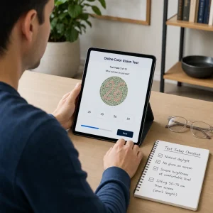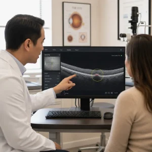
A visual field test is simpler than it sounds. It checks how well you see straight ahead and off to the sides while you keep your eyes fixed on one central target. If you have been told you need one, this guide explains the names, the numbers, and what the results can and cannot tell you.
What a visual field test is in plain English
A visual field test measures your central and peripheral vision without asking you to move your eyes. Perimetry is the clinical name for this kind of testing, and Humphrey visual field testing is one common computerized version used in eye care.
Your visual field is everything you can see when you stare at one point. Think of it like keeping your eyes on a stoplight while still noticing cars, sidewalks, and pedestrians beside you. That wider awareness is your side vision, also called peripheral vision.
This test helps your optometrist look for patterns of vision loss linked to glaucoma, optic nerve disease, retinal disease, stroke, medication toxicity, and some neurological conditions. It does not replace a full eye exam, because doctors read it together with eye pressure, optic nerve findings, OCT imaging, symptoms, and medical history.
Why an eye doctor might order a visual field test
A doctor orders this test when they need to know whether part of your visual field is missing, weaker, or changing over time. Common reasons include glaucoma screening or monitoring, unexplained side-vision loss, optic nerve concerns, droopy eyelids, medication monitoring such as hydroxychloroquine use, and functional concerns related to daily activities.
The symptoms that lead to testing are usually practical, not technical. People describe bumping into things, missing cars or cyclists on one side, trouble in crowds, patchy blur, uneven vision between the eyes, or being told their optic nerve looks suspicious during an eye exam.
This test can raise concern for neurological problems, but it does not diagnose multiple sclerosis, an aneurysm, or a stroke on its own. Certain patterns may suggest that more urgent medical assessment is needed, which is exactly why the result has to be interpreted in the context of the whole exam.
What the numbers mean: 24-2, 30-2, 10-2 and why the “2” matters
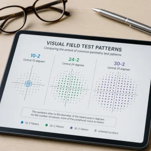
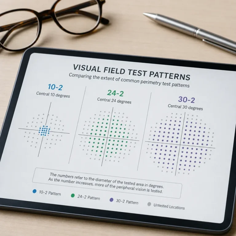
The first number tells you roughly how far from the centre of your gaze the test pattern reaches. In plain terms, a 24-2 visual field test checks the central field out to about 24 degrees, a 30-2 visual field checks a bit farther to about 30 degrees, and a 10-2 visual field test concentrates tightly on the central 10 degrees.
The degrees are about location, not glasses power or eyesight sharpness. If you imagine a circle around the spot you are staring at, 10 degrees is a small centre circle, 24 and 30 degrees spread farther outward, and a 120 degree visual field test is much broader and used for a different purpose.
The “2” in 24-2, 30-2, and 10-2 refers to the way the test grid is offset around the horizontal and vertical midlines. Patients do not need the engineering details, but the short version is that the pattern is designed to sample the field around the centre in a standardized layout rather than choosing random spots.
So what does 24/2 mean on a visual field test? It means the machine is testing the central visual field to roughly 24 degrees from fixation using the standard “-2” grid pattern. What does 30-2 visual field meaning come down to? The same idea, but the tested area extends farther from the centre.
A simple way to picture visual field degrees
A 10-2 test is like zooming in on the centre of your vision. A 24-2 test is a wider central check. A 30-2 visual field gives a bit more spread beyond that. A 120 visual field test looks at a much broader functional field and is usually not the same kind of threshold test used for glaucoma follow-up.
24-2 vs 30-2 vs 10-2 vs 24-2C: which test is used when?
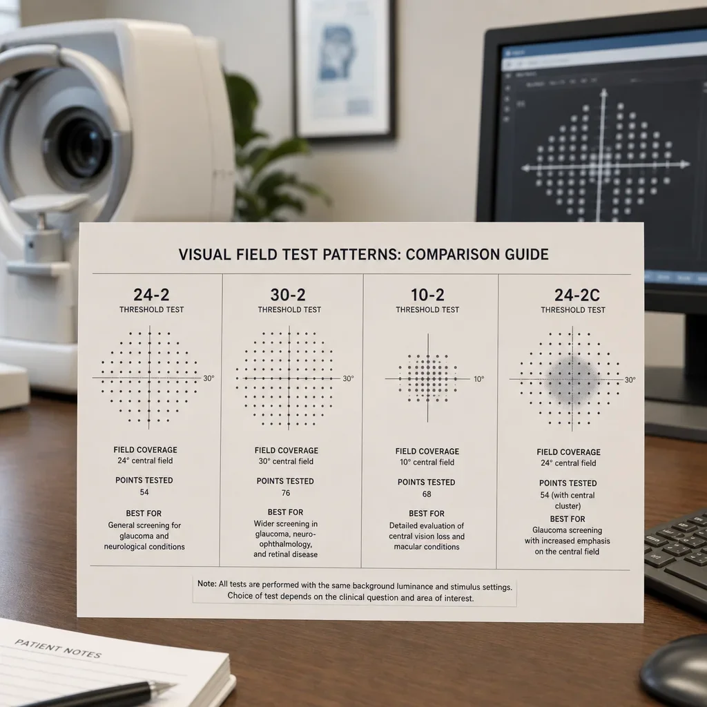
The right protocol depends on where the doctor suspects change and what they need to monitor over time. Symptoms, optic nerve appearance, retinal findings, medication history, and the reason for the visit all matter more than the test name itself.
| Test | Area covered | Common use | Strength | Common limitation |
|---|---|---|---|---|
| 24-2 visual field test | About 24 degrees from centre | Common in glaucoma assessment and follow-up | Good balance of central coverage and practicality | Can miss very small central defects better seen with denser central testing |
| 30-2 visual field test | About 30 degrees from centre | Useful when a wider central field pattern is needed, including some neuro-ophthalmic concerns | Broader central field coverage | Takes longer than tighter patterns |
| 10-2 visual field test | Central 10 degrees | Helpful for central or paracentral defects, some macular concerns, and some medication monitoring | More detailed look near fixation | Too narrow if the concern is farther out in the field |
| 24-2C visual field | Roughly 24-degree pattern with more central sampling | Used when extra central detail is useful while keeping a 24-2 style layout | Better central emphasis than standard 24-2 | Availability and protocol choice vary by clinic and device |
A Humphrey 24-2 visual field test is commonly used when glaucoma is suspected or being monitored. It gives a structured look at the central field area where glaucoma damage can show up, even when patients still feel their vision is normal.
A 30-2 visual field test is often chosen when the doctor wants a wider central map. That can be useful for some optic nerve and neurological patterns, because problems related to the brain or visual pathways may not follow the same pattern as glaucoma.
A 10-2 Humphrey visual field is used when the centre matters most. If someone has central blur, paracentral symptoms, macular concerns, or medication monitoring that needs tighter central testing, 10-2 perimetry gives a closer look at that small area.
The difference between 24-2 and 24-2C is central emphasis. A 24-2C visual field keeps the general 24-2 concept but adds more sampling in the centre, which can help when early central defects are a concern. Exact implementation can differ by platform, so your doctor will usually describe why that protocol was chosen rather than focusing on the device language alone.
Here is the beginner version of the decision guide. Tunnel-vision or wider pattern concerns may push the choice toward a broader field strategy. Central blur or paracentral concerns may lead to a 10-2 visual field. Glaucoma follow-up often uses 24-2, while some cases benefit from 24-2C when central detail matters more.
What is Humphrey visual field testing?
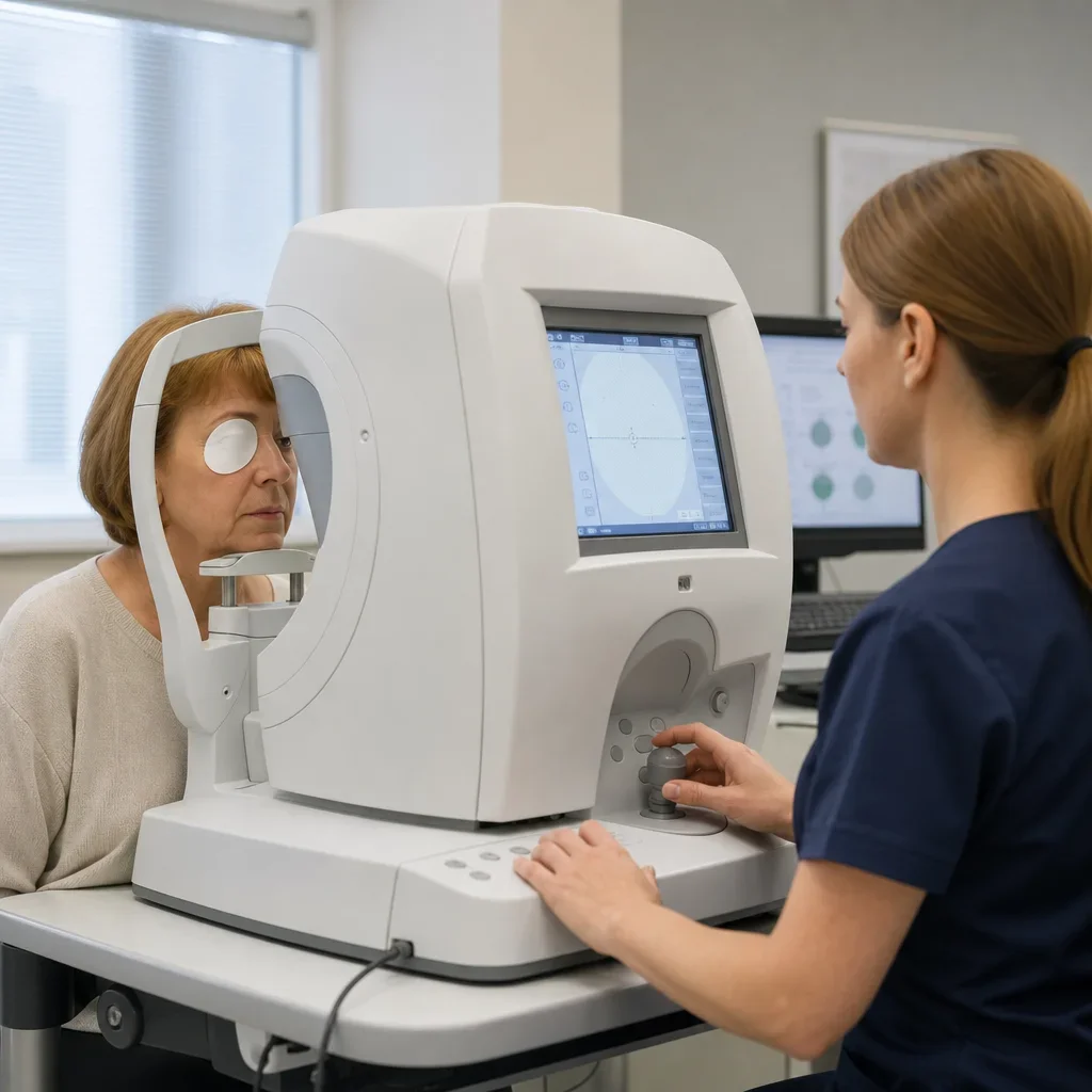
Humphrey visual field, often shortened to HVF, is a common automated form of static perimetry. “Static” means the machine shows lights in fixed locations and changes how bright they are to measure how sensitive different parts of your vision are.
When people say 24 2 HVF or HVF 30-2, they usually mean a Humphrey machine running that specific test pattern. The printout then shows maps and summary indices your optometrist uses to judge whether any changes look real, repeatable, and clinically meaningful.
Static perimetry is different from kinetic perimetry. In static testing, lights appear in set points. In kinetic testing, a target moves from the outside inward until it is seen. Both have a place, but computerized Humphrey-style testing is the format many patients see for glaucoma and related monitoring.
What is a 120 degree visual field test and how is it different?
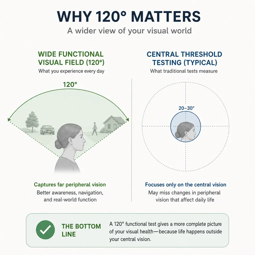
A 120 degree visual field test is broader and more functional than a standard glaucoma threshold test like 24-2 or 30-2. The idea is to assess a wide area of side vision, often for practical function rather than fine threshold mapping of glaucoma change.
You may also hear about a 120 point Esterman test. At a high level, Esterman-style testing is used for functional visual field assessment and may come up in disability or driving-related contexts, but protocols can vary by device, clinic, and the purpose of the test.
This is why a 120 visual field test is not interchangeable with a 24-2 visual field test. One is aimed more at broad functional field screening, while the other is built to measure sensitivity at standardized points in the central field for medical monitoring.
What does a 20 degree visual field mean?
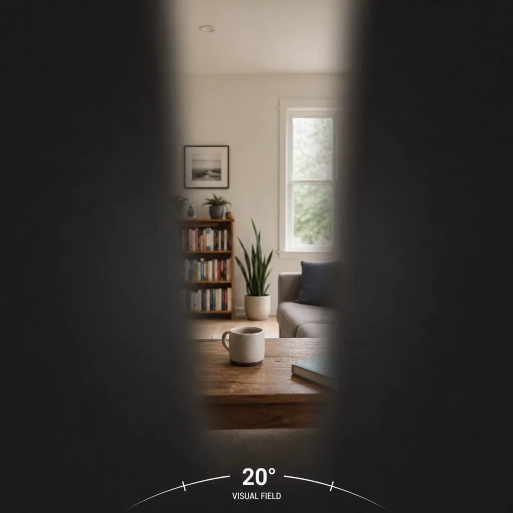
A 20 degree visual field means vision is restricted to a narrow central area. In everyday terms, it can feel like seeing through a tube or tunnel, with much less awareness of what is happening to the sides.
A 20 degree field of vision can make walking in crowds, noticing obstacles, or spotting cars and cyclists from the side much harder. The cause can vary widely, so the number describes the amount of field left, not the reason it happened.
Legal blindness and driving standards are jurisdiction-specific, and the rules can involve field size, visual acuity, or both. That is why we do not tell patients to judge legal status from one printout alone. A proper eye exam and the relevant Ontario standard matter.
What happens during the test and how to prepare
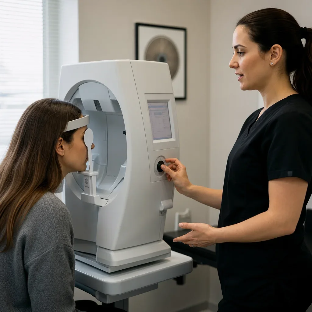
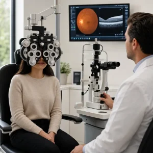
The test is straightforward. One eye is covered, your chin rests on the machine, you stare at one centre target, and you press a button whenever you notice small lights appear.
You are usually told to keep looking straight ahead the whole time. That instruction matters because the machine is testing what you can notice in your side vision while your gaze stays fixed, not how quickly you can search around for lights.
You may wear a trial lens or your glasses may be reviewed depending on the setup and your prescription. In many clinics, the proper correction for the test is provided if needed, because blurry focus can make the result less reliable.
Dilation is not usually needed for the field test itself. If your doctor wants a broader eye exam the same day, dilation may still be recommended for the retinal or optic nerve assessment, but that is separate from the field machine.
The best preparation is practical. Bring your current glasses, arrive rested, blink normally, mention dry eye, and ask for a short pause if you feel tired. Missing some lights is expected, so do not start guessing or chasing lights with your eyes.
How long a visual field test takes
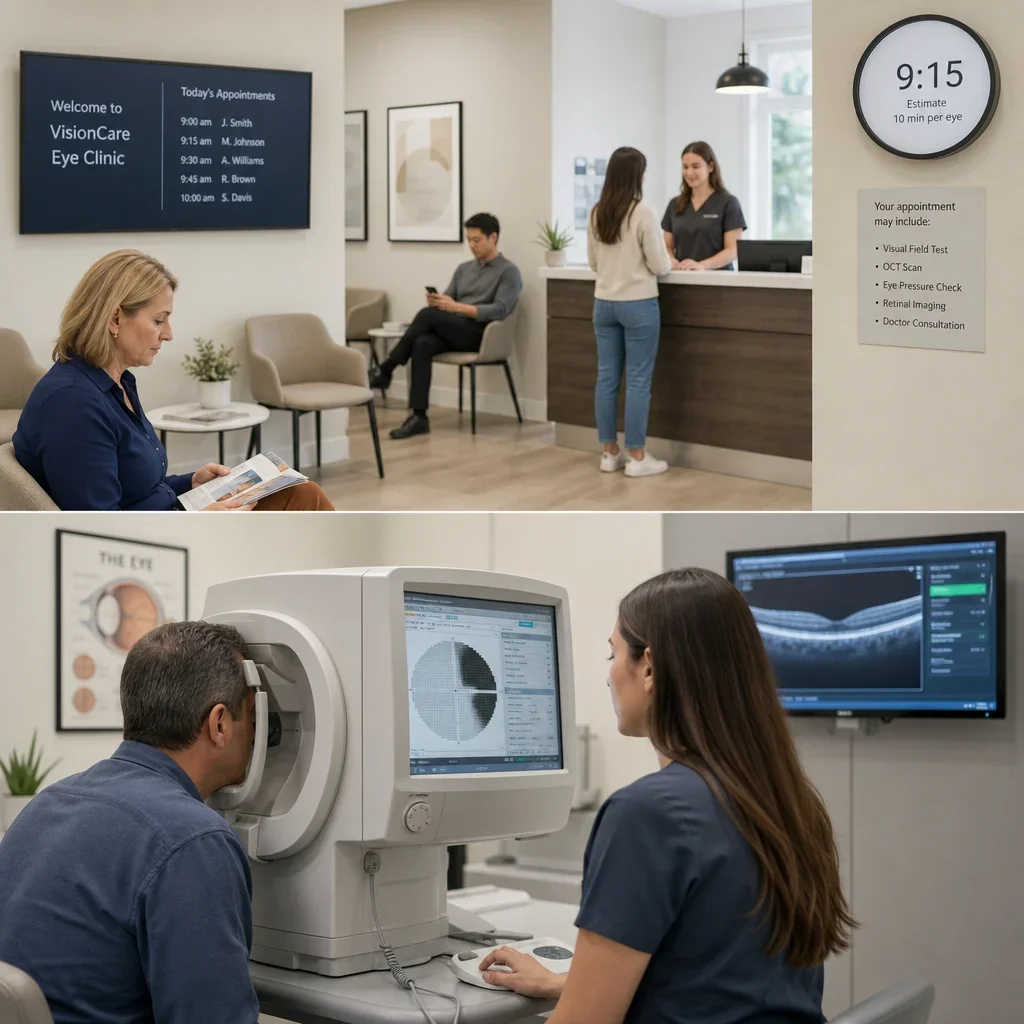
Most visual field tests take about 5 to 10 minutes per eye, and the test portion is often about 15 to 20 minutes total depending on the protocol and how steady the responses are.
A 30-2 visual field test can take longer than a tighter pattern because it samples a larger area. The full appointment may also be longer if the field test is combined with a comprehensive eye exam, pressure check, OCT, or retinal imaging.
Repeat testing is sometimes needed when reliability is poor. That does not mean the patient failed. It usually means the machine detected too much eye movement, fatigue, blur, or inconsistent responses to trust the result fully.
How to read visual field test results without getting lost in jargon

The printout usually has four parts patients notice first: a grayscale map, a table of numbers, deviation plots, and summary scores. The grayscale gives a quick visual impression, but doctors do not diagnose from that picture alone because darker areas can look dramatic without proving a stable defect.
The numerical threshold values show how sensitive different test points were. Higher sensitivity means dimmer lights were still detected, while lower sensitivity means brighter lights were needed at that location.
Total deviation compares your results with age-matched normal expectations at each point. Pattern deviation tries to highlight localized defects by adjusting for any overall depression, which can matter when blur, cataract, or small pupils affect the whole field.
Fixation losses, false positives, and false negatives are reliability checks. Fixation losses suggest you may not have stayed centred consistently. False positives suggest the button was pressed when no light was shown. False negatives suggest missed responses to lights that should have been visible based on nearby results.
Mean deviation, or MD, is a broad summary of overall field sensitivity compared with age-matched norms. Pattern standard deviation, or PSD, summarizes how uneven the field is, and visual field index, or VFI, is another summary number often used to follow glaucoma over time.
One abnormal result is not enough to self-diagnose glaucoma, stroke, or brain disease from an online PDF. Doctors look for repeatable patterns and compare the field with symptoms, eye pressure, optic nerve appearance, OCT scans, retinal findings, and your medical history before making clinical decisions.
Common visual field patterns doctors look for
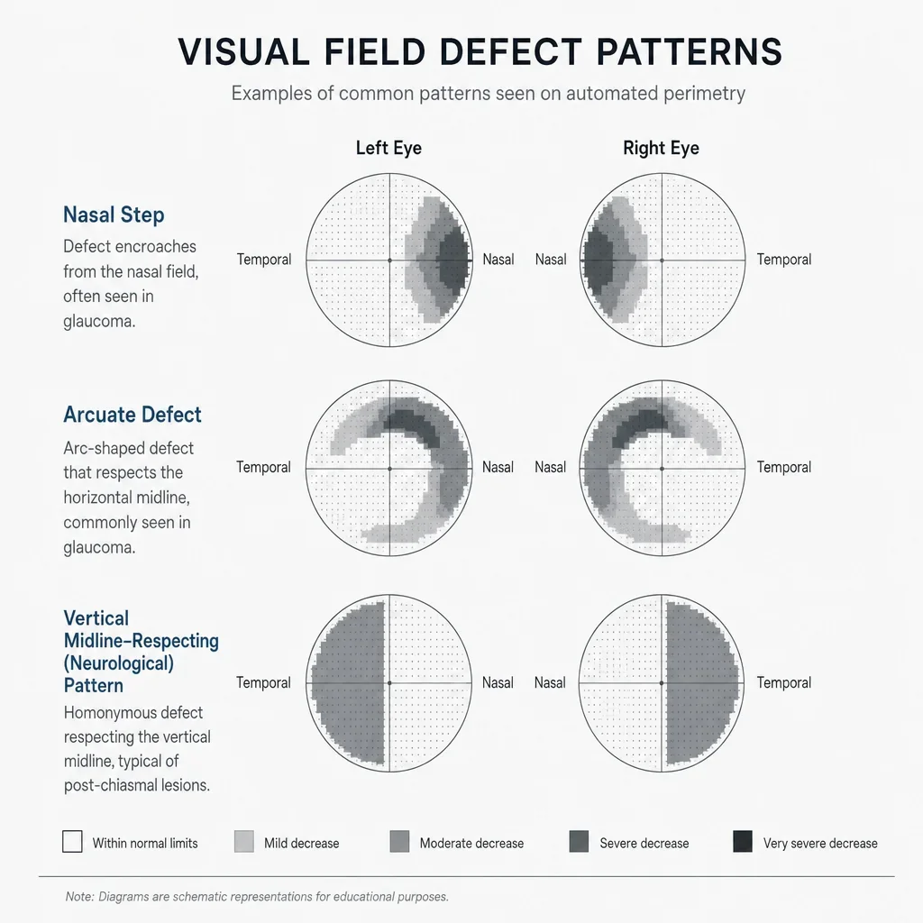
The test can show localized or widespread missing areas called defects. You may hear the word scotoma, which simply means an area where sensitivity is reduced or missing compared with the surrounding field.
Some glaucoma-related patterns have names like nasal step, arcuate defect, and paracentral defect. These terms describe where the loss sits and how it follows the nerve fibre layout of the eye, which is one reason the pattern matters more than one dark patch on a grayscale image.
Neurological patterns can look different. At a high level, glaucoma-related defects often follow the horizontal arrangement of retinal nerve fibres, while some neurological defects may respect the vertical midline, which can point the clinician toward a different part of the visual pathway.
This is also why protocol choice matters. A 24-2 visual field test may be appropriate for broad glaucoma monitoring, while a 10-2 visual field gives a better look when the suspected change is very close to the centre.
What can affect accuracy: tiredness, dry eye, cataracts and other troubleshooting
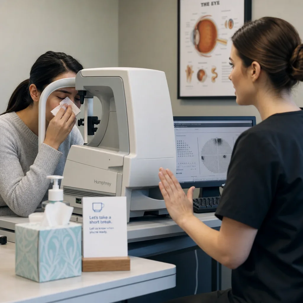
Visual field tests are sensitive to patient comfort and attention. Fatigue, anxiety, dry eye, droopy lids, refractive blur, cataracts, poor fixation, and even fogging or discomfort can make a result look worse than the underlying eye condition really is.
Retesting is common and does not mean you did something wrong. Visual field testing has a learning curve, and many first-time patients do better once they know the rhythm of staring straight ahead and responding without overthinking.
The best way to improve reliability is simple. Tell the clinic if your eyes feel dry, if you are unusually tired, or if you need a short break. Good technicians and doctors would rather pause than collect a poor-quality test that has to be repeated later.
Doctors also trust trends more than isolated results. A field that is stable across repeat visits usually means more than one strange printout taken on a rushed or uncomfortable day.
Visual field tests, glaucoma, and when worsening may show up
Glaucoma often affects peripheral vision first, which is why many people do not notice symptoms early. A patient can feel fine while the test shows subtle side-vision change, especially when the loss develops slowly.
Central vision may stay better until later in some cases, but not always. Modern glaucoma care also pays close attention to early central and paracentral loss, which is one reason some patients are followed with 10-2 testing or central-enhanced patterns like 24-2C.
Worsening is judged over time, not from one number in isolation. Doctors compare repeat fields, optic nerve findings, OCT measurements, eye pressure trends, symptoms, and treatment history before deciding whether glaucoma looks stable or progressive.
Driving, legal blindness, and failing a field vision test
An abnormal field test does not automatically mean you cannot drive. Fitness to drive depends on the amount of loss, where the loss sits, why it is there, whether it is repeatable, and the rules set by the relevant licensing authority.
The phrase “failed a visual field test” can be misleading. Sometimes it means the field was medically abnormal. Other times it means the test was unreliable because of fixation losses, fatigue, or poor concentration. Those are very different situations.
Driving standards and legal blindness definitions vary by province, jurisdiction, and agency. If driving is the issue, the safest next step is a full eye exam and guidance based on the current Ontario standard rather than internet summaries or a single field printout.
When to book an eye exam in Waterloo
Book a comprehensive eye exam if you have been referred for a visual field test, have glaucoma concerns, notice side-vision changes, or want help understanding old Humphrey results. A field test is most useful when it is interpreted alongside the rest of your eye health assessment.
At Premier Optical in Waterloo, patients can book an eye exam with licensed optometrists and get help deciding whether visual field testing, OCT imaging, or other follow-up is appropriate. Direct insurance billing may be available for eligible services, which can make the visit simpler to manage.
If you already have a printout that says 24-2, 30-2, 10-2, or 24-2C, bring it with you. The most useful next step is not guessing from the numbers. It is having the result reviewed in the context of your symptoms, your optic nerve, and your full exam.
FAQ
What is a visual field test?
A visual field test checks how well you see in the centre and to the sides while you stare at one fixed point. It is a form of perimetry and is commonly used in glaucoma and optic nerve assessment.
What does 24-2 mean on a visual field test?
It means the test pattern examines the central field out to about 24 degrees from where you are looking, using the standard “-2” grid layout.
What is the difference between 24-2 and 30-2 visual field tests?
A 30-2 visual field reaches farther from the centre, to about 30 degrees, while a 24-2 focuses on about 24 degrees. The doctor chooses between them based on the pattern they want to assess.
What is the difference between 10-2 and 24-2 visual field tests?
A 10-2 test concentrates on the central 10 degrees, so it is better for fine central detail. A 24-2 covers a wider central area and is commonly used in glaucoma monitoring.
What is a 24-2C visual field test?
A 24-2C visual field is a central-enhanced version of the 24-2 concept. It adds more central sampling while keeping a roughly 24-degree pattern.
What is a 120 degree visual field test?
It is a broader functional field assessment, not the same as a standard glaucoma threshold test like 24-2 or 30-2. It may be used for wider functional questions, including some driving-related assessments.
How long does a visual field test take?
Many tests take about 5 to 10 minutes per eye, though timing varies by protocol, patient comfort, and test reliability.
Do they dilate your eyes for a visual field test?
Usually not for the field test itself. Dilation may still be part of the full eye exam if your optometrist needs a better view inside the eye.
Do you wear glasses for a visual field test?
Sometimes a trial lens is used instead of your regular glasses, depending on the machine and your prescription. The goal is to give the test the right focus.
What should you not do before a visual field test?
Do not arrive exhausted if you can avoid it, and do not try to hunt for lights by moving your eyes around. Staying relaxed, blinking normally, and looking straight ahead gives a more reliable result.
How do I read my visual field test results?
Start with reliability, then pattern, then summary scores like MD, PSD, and VFI. The result still needs to be interpreted with your eye exam, because one printout alone is not a diagnosis.
What is a normal visual field test result?
A normal result shows sensitivity within the expected range for your age without a repeatable defect pattern. Your optometrist judges normality using the full printout, test reliability, and the rest of the exam.
Can I drive if I fail a field vision test?
Not every abnormal result means you cannot drive. The answer depends on the reason for the test, how reliable it was, how much field is affected, and the official standard that applies to you.
Can an eye doctor detect multiple sclerosis from a visual field test?
No test like this diagnoses MS by itself. A visual field can show patterns that raise concern for optic nerve or neurological problems, but diagnosis requires broader medical assessment.