A funduscopic examination is simply a close look at the back of the eye. In a Waterloo eye exam, that can mean an ophthalmoscopy exam, a dilated retinal check, or retinal imaging, depending on your symptoms and what the optometrist needs to assess.
What a funduscopic examination is, in plain English
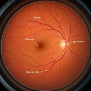
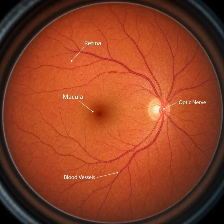
A funduscopic examination looks at the inside back of your eye, called the fundus oculi. That area includes the retina, macula, optic nerve, and retinal blood vessels.
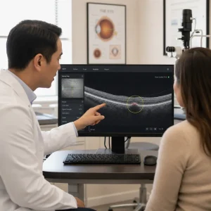
Fundoscopy examination and ophthalmoscopy exam mean the act of examining that area. A fundus photo eye test means taking pictures of it, while OCT creates cross-sectional images of retinal layers rather than a surface photo.
An ocular fundus exam may be part of a comprehensive eye exam, or it may be done because of diabetes, high blood pressure, glaucoma concerns, flashes, floaters, headaches, or blurred vision. In a community clinic, the same visit may include direct viewing, a slit-lamp retinal exam, dilation, and imaging if needed.
What parts of the eye this exam checks
An eye exam fundus check looks at structures that affect both sight and eye health. The retina senses light, the macula handles sharp central vision, the optic nerve carries signals to the brain, the blood vessels reflect circulation, and the peripheral retina is where tears or detachments can start.
The optic fundus examination matters because serious changes can begin at the back of the eye before you notice symptoms. That is why a fundus check up can be useful even when vision seems fine.
Why a fundus exam is done and who should get one
A fundus exam is done to check retinal and optic nerve health, either routinely or because symptoms raise concern. Common reasons include diabetes follow-up, high blood pressure concerns, glaucoma monitoring, new flashes or floaters, sudden blur, headaches with visual symptoms, and retinal risk checks.
The right timing depends on age, symptoms, medical history, family history, and the optometrist’s findings, not one universal schedule. Some people need only routine monitoring, while others need prompt review because the symptom pattern is new or sudden.
A practical rule is simple. Gradual blur or a routine diabetes review usually fits a regular appointment, but sudden flashes, a shower of new floaters, a curtain or shadow in vision, or a sudden drop in sight needs same-day assessment.
What conditions a funduscopic examination can detect
A funduscopic examination can show signs of retinal disease, optic nerve disease, and blood vessel changes inside the eye. The most common findings patients ask about are diabetic retinopathy, hypertensive retinal changes, glaucoma-related optic nerve cupping, age-related macular degeneration, retinal tears, retinal detachment, and optic nerve swelling such as papilledema.
An ophthalmoscopy test can also show bleeding, swelling, drusen, pigment changes, or vessel abnormalities that need follow-up. Those findings can raise suspicion for an eye condition, but they do not replace bloodwork, brain imaging, or a full medical assessment when systemic disease is a concern.
Some systemic issues can leave signs at the back of the eye. High blood pressure may affect retinal vessels, diabetes may affect capillaries, and cholesterol can sometimes be associated with visible ocular findings, but an eye exam alone does not diagnose high cholesterol.
| Finding seen on fundus exam | What it may suggest | Usual urgency |
|---|---|---|
| Microaneurysms or retinal bleeding | Diabetic retinopathy or other vascular change | Prompt follow-up |
| Vessel narrowing or crossing changes | Blood pressure-related retinal change | Routine or prompt follow-up |
| Optic nerve cupping | Glaucoma suspicion or monitoring need | Routine or prompt follow-up |
| Drusen at the macula | Age-related macular degeneration changes | Routine or prompt follow-up |
| Retinal tear or hole | Risk of retinal detachment | Urgent |
| Optic nerve swelling | Papilledema or other optic nerve issue | Urgent |
First signs and urgent red flags patients should not ignore

Some retinal and optic nerve symptoms should not wait for a routine booking. Sudden flashes of light, a shower of new floaters, a curtain or shadow over vision, sudden loss of part of vision, sudden major blur in one eye, or new central distortion can be urgent.
A retinal tear may start with flashes and floaters, and a retinal detachment may add a shadow, curtain, or missing area of vision. Headache with nausea and new visual symptoms can also need urgent assessment if optic nerve or pressure-related causes are being considered.
Seek urgent eye assessment today if you have sudden flashes, many new floaters, curtain-like vision, sudden one-eye blur, or sudden vision loss.
Long-standing mild blur is different from a sudden change. Chronic prescription blur can often wait for a regular eye exam, but acute change is treated differently because timing matters.
What happens during the appointment, minute by minute
The retinal part is usually brief, but the full visit takes longer because history, vision testing, and any imaging come first. A comprehensive eye exam appointment commonly runs about 20–60+ minutes, while the actual retinal viewing or fundus camera eye test may take only a few minutes.
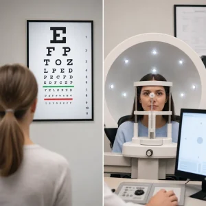
The visit usually starts with your symptoms, medical history, medications, glasses or contact lens use, and a vision check. Preliminary tests may come next, followed by retinal imaging, dilation if needed, the retinal examination itself, and a review of findings.
The physical experience is usually bright light, short instructions to look in certain directions, and no pain. If dilation is used, the drops often need about 15–30 minutes to work before the fullest retinal view is possible.
Do you need dilation? What the drops do and how long they last

Dilation is not always required, but it may be recommended when the optometrist needs a wider or clearer view of the retina. It is especially useful when symptoms involve the peripheral retina, when retinal tears are a concern, or when a fuller diabetic or optic nerve assessment is needed.
Dilating drops make the pupil larger so more of the back of the eye can be seen. Temporary effects commonly last several hours, often about 2–6 hours, though some people notice blur or light sensitivity longer depending on the drops used and the individual response.
The usual after-effects are light sensitivity and blurry near vision. Bringing sunglasses helps, and work or school is still possible for many people, but reading and screen comfort may be worse until the drops wear off.
Driving after dilation is not something we guarantee. Your ability depends on the drops used, your light sensitivity, your vision, and how you feel afterward, so follow the optometrist’s advice on the day of the exam.
Contact lenses may need to be removed for parts of the visit, especially if drops or imaging are planned. If you wear contacts, bring your case and glasses so you have options after the exam.
Dilation aftercare checklist
- Bring sunglasses for the trip home.
- Expect bright light sensitivity for a few hours.
- Expect near blur if your pupils are dilated.
- Bring glasses and a contact lens case.
- Ask before driving yourself home.
Is a fundus exam painful or risky?
A fundus exam is typically not painful. The most common sensations are bright light, mild temporary discomfort from glare, and temporary blur if dilating drops are used.
Direct fundoscopy, slit-lamp retinal viewing, and a fundus photo eye test are non-invasive. That means nothing touches the back of your eye during the actual viewing or photography.
Side effects are usually limited to short-term light sensitivity or blur after dilation. Rare reactions to drops can happen, which is one reason your history and symptoms are reviewed before the exam.
Types of fundus examination: direct, indirect, slit-lamp, photos, widefield, and OCT
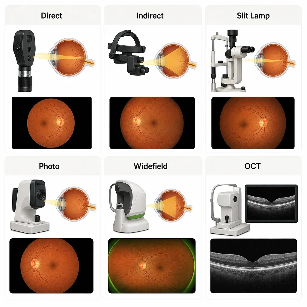
Different tests answer different questions, and no single method replaces every other one. Direct fundoscopy, indirect viewing, slit-lamp retinal examination, fundus photography, widefield imaging, and OCT each have different strengths and limits.
| Test type | What it is | Best for | Dilation may be needed | Permanent image? | Main limitation |
|---|---|---|---|---|---|
| Direct fundoscopy | Handheld lighted view into the eye | Quick optic nerve and central retina view | Sometimes | No | Narrower view |
| Indirect fundoscopy | Wider retinal view using special optics | Peripheral retina and tear risk | Often | No | Not a stored photo by itself |
| Slit-lamp retinal exam | Doctor examines retina through microscope optics | Detailed clinical examination | Sometimes | No | Depends on view and cooperation |
| Fundus photography | Surface photos of retina | Baseline documentation and comparison over time | Sometimes | Yes | A photo can miss some findings outside the captured view |
| Widefield retinal photography | Broader retinal photos | More peripheral documentation | Sometimes | Yes | Still not identical to a full dynamic dilated exam |
| OCT | Cross-sectional retinal scan | Macula and optic nerve layer analysis | Not always | Yes | Shows layers, not the whole retinal surface in one image |
The key consumer point is simple. Direct fundoscopy and slit-lamp viewing let the doctor examine the eye live, while imaging creates a record that can be compared over time. Photos are helpful, but photography alone can miss problems a dilated exam can catch.
Fundus photography vs OCT vs a dilated eye exam
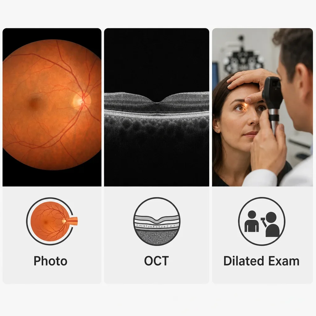
Fundus photography shows the retinal surface, OCT shows cross-sectional retinal layers, and a dilated exam lets the doctor assess the eye directly and dynamically. They are not interchangeable, even though they are often used together.
A fundus camera eye test is useful for documenting diabetic changes, macular appearance, optic nerve appearance, or a baseline for future comparison. OCT is especially useful when the concern involves the macula or optic nerve layers, such as swelling, thinning, or glaucoma-related change.
A dilated eye exam is often the better choice when symptoms suggest a peripheral retinal problem, including flashes, floaters, or a possible retinal tear. There is no universal best test because the right choice depends on symptoms, risk factors, and what needs to be ruled out.
Retinal imaging is helpful, but it is not automatically mandatory in every comprehensive eye exam. Ask whether it is included, optional, recommended for your risk level, or advised because of a specific finding.
What a normal fundus exam result looks like
A normal fundoscopic exam means the retina, macula, optic nerve, and vessels look healthy on that visit. In patient language, that usually means no retinal tears, no swelling, no bleeding, no diabetic retinopathy seen, and no concerning optic nerve change identified.
Normal fundoscopic exam findings may be described with wording such as within normal limits, healthy optic nerve appearance, macula clear, retinal vessels normal, or no holes or tears seen. A normal fundus examination report can still be followed by routine monitoring if age, diabetes, family history, or glaucoma risk factors are present.
What abnormal findings can mean
An abnormal fundus report means something was seen that needs interpretation in context, not that a diagnosis is final from one image or one phrase. Common report terms can sound alarming, but they are often the start of a next-step plan rather than a final answer.
| Report term | Plain-English meaning | What usually happens next |
|---|---|---|
| Retinal hemorrhage | Bleeding seen in the retina | Cause assessed, follow-up or referral arranged |
| Drusen | Deposits linked with macular aging changes | Monitoring or macular assessment |
| Cupping | Optic nerve shape change associated with glaucoma risk | Glaucoma testing or monitoring |
| Retinal tear or hole | Weak or open area in peripheral retina | Urgent retinal assessment |
| Papilledema | Optic nerve swelling | Urgent medical or specialist assessment |
| Diabetic changes | Diabetes-related retinal findings | Diabetic eye follow-up plan |
| Vessel narrowing | Blood vessel change that may relate to blood pressure | Medical correlation and eye follow-up |
The safest way to read any fundoscopy report is to pair the wording with the clinical explanation given at the visit. One sign can have more than one cause, which is why history, symptoms, pressure testing, vision testing, and imaging may all matter together.
Fundus examination cost in Ontario: what patients may pay
The first cost question is really three separate questions: the eye exam fee, whether a dilated retinal exam is included, and whether retinal imaging is extra. In Ontario, private-pay comprehensive eye exams commonly fall around C$120–C$250, depending on the clinic, the visit type, and what is included.
Fundus examination cost can change based on whether the retinal check is simply part of the doctor’s exam or whether extra imaging is offered as an add-on. Some clinics charge a separate fee for fundus photography or OCT, while others bundle some testing into selected exam types.
The most practical question to ask is not just the fundus examination test price. Ask whether dilation is included, whether a fundus camera eye test is optional or recommended, whether OCT would be extra, and whether the testing is being done for routine screening or a medical concern.
Insurance, OHIP, and direct billing: what to ask before you book
Public coverage and private coverage are not the same thing. In Ontario, some eye exams may be publicly covered for eligible groups or medically necessary reasons, while many routine exams are private-pay.
Private insurance may cover part or all of a comprehensive eye exam, and some plans also cover retinal imaging, but coverage rules depend on the insurer and the plan wording. If you are comparing the cost of fundus eye test options, ask if the imaging is claimable, whether it is considered part of the exam, and what receipt wording your insurer needs.
Direct billing can make the visit simpler because the clinic can often submit to participating insurers at the time of service. Bring your insurance card, photo ID, and any referral or previous prescription details if you have them.
When to book a routine eye exam vs seek urgent same-day care
Routine booking fits checkups, gradual vision changes, diabetes follow-up, and ongoing glaucoma monitoring. Same-day care fits sudden flashes, many new floaters, curtain-like vision, sudden one-eye blur, eye trauma, or sudden vision loss.
If you are in Waterloo or Kitchener and want a thorough eye health check, book a comprehensive exam with a licensed optometrist and ask whether retinal imaging, dilation, or both are recommended for your visit. If you have urgent retinal red flags, call immediately for guidance or seek same-day urgent assessment.
FAQ
What is a funduscopic examination?
It is an exam of the back of the eye, including the retina, macula, optic nerve, and blood vessels.
Is a fundus exam painful?
It is usually not painful. Bright light and temporary blur after dilation are the most common discomforts.
How long does a fundus test take?
The retinal viewing itself may take only a few minutes, while the full eye exam commonly takes about 20–60+ minutes depending on testing and dilation.
Do I need dilation for a fundus exam?
Not always. Dilation may be recommended when a wider retinal view is needed, especially for peripheral retina concerns or a more complete assessment.
How long do dilation effects last?
They commonly last several hours, often about 2–6 hours, though some people notice effects longer.
What conditions can a fundus exam detect?
It can show signs of diabetic retinopathy, blood pressure-related retinal changes, glaucoma-related optic nerve changes, macular degeneration, retinal tears, retinal detachment, and optic nerve swelling.
What is the difference between fundoscopy and fundus photography?
Fundoscopy is the doctor examining the back of the eye. Fundus photography is taking pictures of it for documentation and comparison.
What is the difference between fundus photography and OCT?
Fundus photography shows the retinal surface. OCT shows cross-sectional retinal layers.
Is a fundus exam the same as a regular eye test?
It can be one part of a comprehensive eye exam, but it is not the whole visit by itself.
Can an eye exam show glaucoma?
An eye exam can show optic nerve changes that raise concern for glaucoma and can support glaucoma monitoring, but diagnosis uses the full clinical picture.
Can an eye exam show high cholesterol?
It may show certain eye findings that raise suspicion, but it does not diagnose high cholesterol on its own.
What is a normal fundus exam result?
It usually means healthy retinal appearance, healthy optic nerve appearance, normal vessels, and no signs of tears, bleeding, swelling, or diabetic changes on that visit.
What should I expect after ophthalmoscopy?
If no dilation is used, usually very little. If dilation is used, expect light sensitivity and blurry near vision for a few hours.
Can I drive after my pupils are dilated?
Some people can, some should not. It depends on the drops, your vision, your light sensitivity, and the optometrist’s advice.
Do I need to remove contact lenses for a fundus exam or fundus photo?
Sometimes. Bring your glasses and a contact lens case in case removal is needed.
How much does a fundus examination cost in Ontario?
The answer depends on whether you mean the eye exam itself, retinal imaging, or both. Private-pay comprehensive eye exams commonly run about C$120–C$250 in Ontario, and imaging may be extra depending on the clinic.
Is fundus photography covered by insurance or OHIP?
Sometimes by private insurance, depending on your plan. OHIP and other public coverage depend on eligibility and medical necessity rules, so confirm with the clinic and your insurer.
When should flashes and floaters be treated as urgent?
Treat them as urgent if they are sudden, numerous, paired with flashes, or followed by a curtain, shadow, or missing part of vision.