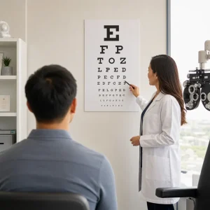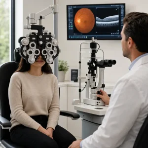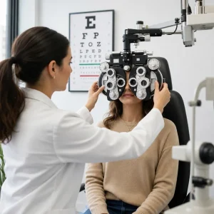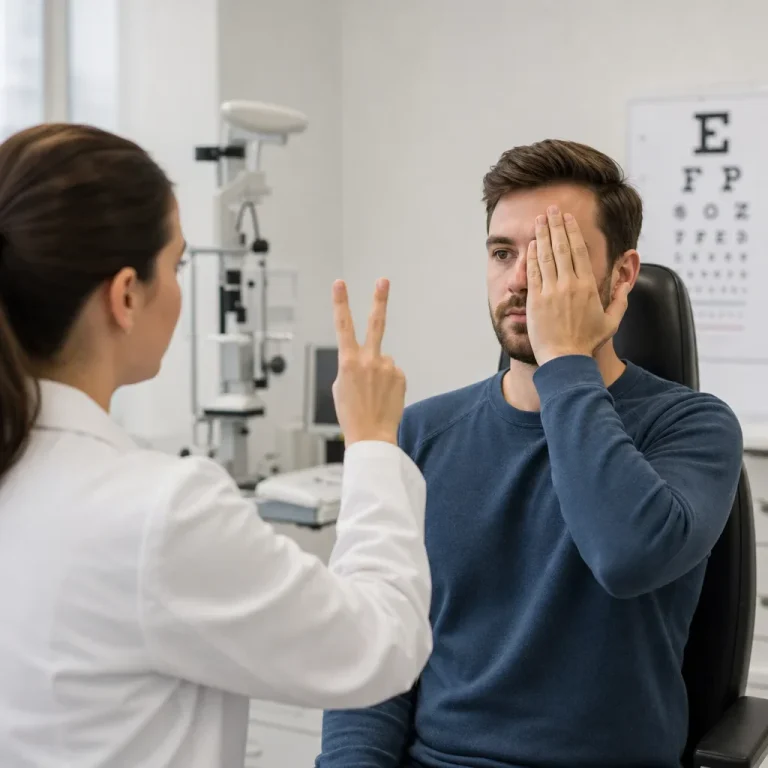
An eye test for peripheral vision checks how well you see to the sides while you keep looking straight ahead. It is different from a standard eye chart because it measures your visual field, not just how clearly you see letters in the centre.
What Is an Eye Test for Peripheral Vision?
A field test for eyes measures how much you can detect above, below, and to each side without moving your gaze. Eye doctors may call this a peripheral eye exam, a visual field test, or perimetry.
Peripheral vision is the side vision you use for walking through crowds, noticing motion, spotting stairs, and staying aware while driving. Central vision is the sharp vision you use for reading, faces, and fine detail.
A field test eye exam matters because people can read the chart well and still miss areas in the side vision. That is why an eye exam peripheral vision test can be part of screening, diagnosis, or follow-up.
Why You Might Need This Test
A visual field test is used to screen for problems, help diagnose them, or monitor changes over time. Common reasons include glaucoma suspicion, optic nerve concerns, retinal disease, droopy eyelids that block vision, unexplained headaches, and symptoms such as bumping into things or missing objects off to one side.
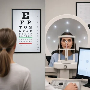
You can have field loss with 20/20 vision because visual acuity and visual field measure different things. The eye chart checks detail in the centre. A test to check peripheral vision looks at how much of the surrounding field you detect.
This test can suggest whether a problem may involve the retina, optic nerve, eyelid, or visual pathways in the brain, but it does not diagnose every cause by itself. If the pattern is concerning, the next step may include a full eye exam, optic nerve assessment, imaging, dilation, or referral.
How Peripheral Vision Is Tested at an Eye Exam
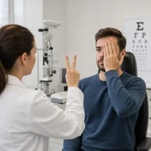
The first screening method is often the confrontation peripheral vision test. You cover one eye, look at a target, and say when you see fingers or a moving target in the side vision. This is a quick way to assess peripheral vision in the exam room.
The more precise method is automated perimetry. You look into a bowl-shaped machine, keep one eye fixed on a central target, and press a button when small lights appear. Automated testing usually takes about 5 to 10 minutes per eye.
One eye is tested at a time because each eye can have a different field pattern. Covering one eye also helps the clinician measure peripheral vision accurately instead of blending what both eyes see together.
Static perimetry uses fixed lights in different spots and brightness levels. Kinetic perimetry uses moving targets that come in from the outer field until you detect them. Goldmann testing is a manual form of kinetic perimetry used for some complex cases or detailed mapping.
A 24-2 visual field test is a common automated pattern used in glaucoma care. It tests points in the central and near-peripheral field in a regular grid rather than mapping the farthest edges of vision.
The test can feel repetitive because the machine presents many faint lights to measure threshold sensitivity, not just whether you can see obvious targets. That repetition is part of how it checks consistency and maps subtle defects.
Confrontation Test vs Automated Visual Field Test vs Goldmann vs Home Checks
| Method | Where used | What you do | Strengths | Limits | Best use |
|---|---|---|---|---|---|
| Confrontation testing | Regular exam room | Look straight ahead and report fingers or movement | Fast, no machine needed | Less sensitive for subtle loss | Quick screening |
| Automated perimetry | Diagnostic testing room | Press a button for small lights in a bowl | More precise and repeatable | Can be tiring, needs focus | Glaucoma and optic nerve monitoring |
| Goldmann/manual kinetic testing | Specialized testing | Respond to moving targets | Helpful for complex or unusual defects | More operator-dependent | Detailed mapping in selected cases |
| Home checks | At home | Compare sides, finger wiggle, symptom tracking | Easy and immediate | Not diagnostic, can miss early disease | Screening only, not a substitute for care |
| Nasal field concern review | Clinic assessment | Formal mapping when one side seems reduced | Better at defining exact defect pattern | Needs professional testing | Clarifying a nasal peripheral vision test concern |
A home or online peripheral vision test at home can be useful for noticing a change, but it cannot assess peripheral visual fields with the same accuracy as office testing. Early glaucoma and subtle neurologic defects can be missed without formal perimetry.
Step by Step: What the Test Feels Like
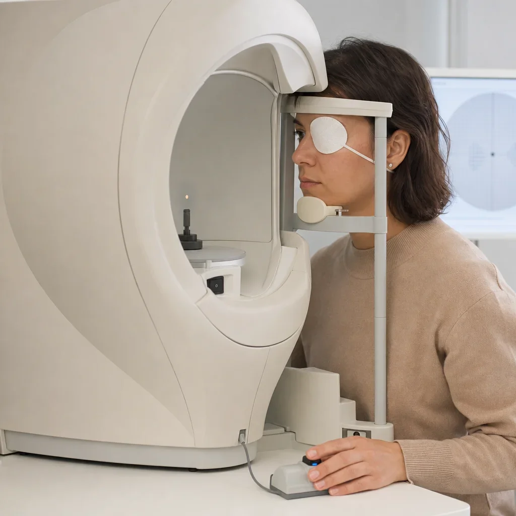
The setup is simple. A staff member seats you at the machine, patches one eye, and aligns your head on a chin rest. Trial lenses may be placed in the device if you need distance correction.
The instructions are to keep looking at the centre and click only when you actually see a light. Some lights are bright and obvious. Others are very dim on purpose.
Blinking is allowed and normal. If your eyes feel dry or you lose focus, a short pause can help the test quality.
The machine also checks how reliable the responses are by tracking fixation losses, false positives, and false negatives. That is one reason a doctor may repeat the test before making conclusions.
How to Prepare for a Visual Field Test
The best way to “pass” is not to game the test. Honest, steady responses produce the most useful result. There is no benefit to clicking wildly or trying to anticipate lights.
Fatigue can affect performance, so getting decent rest helps if you can. Dry eyes, anxiety, and rushing can also make a failed peripheral vision test more likely even when no disease is present.
Bring your current glasses and contact lens details unless the clinic tells you otherwise. Many clinics use your distance correction or trial lenses in the machine instead of your own frames because frame edges can block the field.
Dilation is usually not required for the visual field test itself. It may still be part of the full eye exam if the optometrist needs to examine the retina or optic nerve.
Preparation checklist
- Get enough rest if possible.
- Bring current glasses and contact lens information.
- Ask for the instructions again if anything is unclear.
- Blink normally and mention dry eye or fatigue.
- Do not guess repeatedly or click before you see a light.
- Tell the clinic if you need a short pause.
What a Normal Peripheral Vision Test Result Means
A normal peripheral vision test means you detected the expected pattern of lights for your age and test setup, with good reliability. Doctors look at the shape of the field, missed spots, consistency, and machine reliability markers, not just one number.
A normal printout may include a grayscale map, a blind spot area, deviation plots, and reliability indices. The blind spot is normal because that area corresponds to where the optic nerve exits the eye.
Approximate normal field extent is about 90 to 100 degrees temporally, about 60 degrees nasally, about 60 degrees superiorly, and about 70 to 75 degrees inferiorly. Those are broad anatomy references, not a single pass-fail standard for every machine and patient.
Normal peripheral vision test results also depend on pupil size, focus, attention, eyelid position, and test quality. That is why a doctor may still repeat a normal-looking test if they are watching for trend changes over time.
What Abnormal or Failed Results Can Mean
A failed peripheral eye test does not automatically mean permanent vision loss or a serious disease. It means the result needs interpretation in context of symptoms, exam findings, optic nerve appearance, and sometimes repeat testing.
True abnormal visual field test results can be linked to glaucoma, retinal disease, optic nerve disease, stroke, other neurologic conditions, or even an eyelid physically blocking part of the field. One-eye defects can matter because asymmetry may point to a localized eye or optic nerve issue.
False-fail results are common enough that repeat testing is routine. Fatigue, poor fixation, blinking, dry eyes, misunderstanding instructions, heavy lids, anxiety, or incorrect lens positioning can all affect the map.
What happens next after a failed test
- Repeat the test if reliability was poor.
- Review eye pressure, optic nerve, and the rest of the exam.
- Consider imaging or further testing if clinically indicated.
- Dilate the eyes if the doctor needs a better retinal view.
- Refer for medical or specialist assessment if the pattern suggests a non-routine cause.
If you were told you failed elsewhere and want the result reviewed, booking a full eye exam is the practical next step. At Premier Optical in Waterloo, patients can have an exam, eyewear advice, and in-house service under one roof, with direct insurance billing available where eligible.
Why Visual Field Tests Are Sometimes Repeated
Repeat testing is common because the machine measures both vision and response reliability. A person can press when no light appeared, miss an easy light because they were tired, or look around instead of holding fixation.
Fixation losses mean the eye likely drifted away from the centre target. False positives mean the button was pressed when no stimulus should have been seen. False negatives mean obvious stimuli were missed after sensitivity had already been established.
There is also a learning effect. Many people do better on the second test because they understand the rhythm and know that faint lights are normal. That is why a single abnormal screen is not the same thing as a confirmed diagnosis.
Common Visual Field Patterns Explained in Plain English
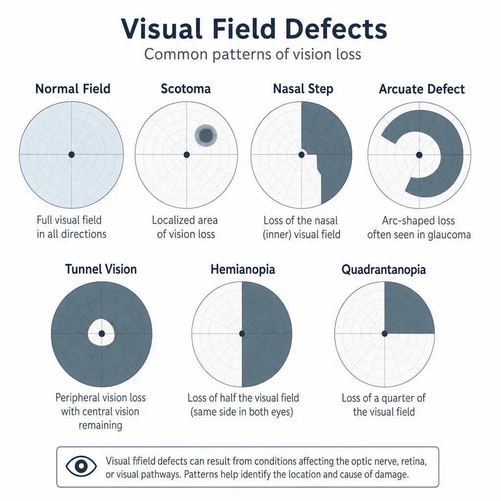
A scotoma is a blind spot or dim area inside the visual field. It can be small or large, central or peripheral.
A nasal step is a defect on the nose side of the field that follows a typical nerve fibre pattern. It is one pattern doctors watch for in glaucoma.
An arcuate defect is a curved area of missing vision that arches from the blind spot area. This can also fit glaucomatous damage patterns.
Tunnel vision means the remaining field is constricted, leaving mainly central vision. Patients may still read an eye chart well and yet struggle badly with navigation.
A hemianopia means half of the visual field is missing. A quadrantanopia means one quarter is missing. Matching left-right losses in both eyes can suggest a problem farther back in the visual pathway rather than in just one eye.
Bitemporal hemianopia affects the outer halves of both visual fields. Homonymous hemianopia affects the same side of the field in both eyes. These patterns can point toward neurologic pathway issues and need medical assessment.
A nasal peripheral vision test concern usually means someone feels reduced awareness on the nose side of one eye’s field. Formal testing helps show whether that pattern is real, repeatable, and more consistent with eyelid blockage, glaucoma, retinal disease, or another cause.
Conditions Linked to Peripheral Vision Loss
Glaucoma is one of the major causes of gradual peripheral field loss. It can damage the optic nerve before a person notices symptoms, which is why visual field testing and optic nerve assessment are often paired.
Retinal problems can also reduce side vision. Retinal detachment may cause flashes, many new floaters, or a curtain-like shadow and needs urgent care. Other retinal disease can create patchy or localized defects.
Optic neuritis and other optic nerve disorders can affect how the field is seen even when the front of the eye looks fairly normal. A visual field test may suggest optic nerve involvement, but it does not diagnose conditions such as multiple sclerosis by itself.
Neurologic causes can include stroke, compression along the visual pathway, or other brain-related conditions. A visual field defect can be one clue, but the pattern has to be matched with symptoms, history, and medical assessment.
Peripheral Vision, Glaucoma, and OCT
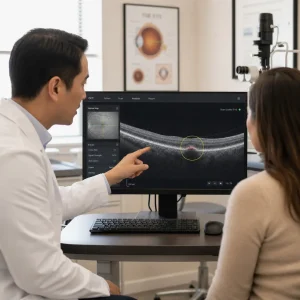
Visual field testing measures function. OCT measures structure, such as the retinal nerve fibre layer and optic nerve anatomy. These tests answer different questions, which is why both may be recommended in glaucoma workups or follow-up.
A person may have structural change before measurable functional loss, or field loss before the pattern is obvious on a basic exam. Looking at both structure and function gives a fuller picture than either test alone.
OCT often does not require dilation, though the overall exam plan can change that. Whether OCT is needed depends on the clinical findings, not on a generic screening rule.
Can You Drive If You Have Peripheral Vision Loss?
Driving safety depends on the cause, severity, stability, and the licensing standards that apply to the driver. Reduced side awareness can affect lane changes, cyclists, pedestrians, and hazard detection.
A field of vision test for driving may be relevant if a person has known field loss or has been advised to follow up after an abnormal result. We would not treat one office test as legal advice, and Ontario licensing questions should be checked against current provincial standards and your eye doctor’s guidance.
If you have new field loss and are unsure whether it is safe to drive, ask directly before getting behind the wheel. Safety decisions should be based on the actual pattern and cause, not on guesswork.
Can You Test Peripheral Vision at Home?
A home check can help you notice a difference, but it cannot replace a professional exam. A simple peripheral vision test with fingers involves covering one eye, looking straight ahead, and comparing when you notice finger movement from each side.
Home checks are limited because your own attention, lighting, distance, and technique change the result. They are screening only, and they can miss early disease or subtle defects.
If one eye seems different from the other, or if your side vision changed suddenly, book an eye exam promptly. If the change is sudden and comes with flashes, floaters, pain, or neurologic symptoms, seek urgent assessment the same day.
Cost, Insurance, and Booking a Peripheral Vision Test in Waterloo
The cost depends on whether the test is part of a comprehensive exam, ordered as an added diagnostic test, or repeated for monitoring. Insurance coverage also varies by plan, provider, and the reason the test was done.
Direct billing may be available for eligible plans, but coverage should be confirmed against your own benefits. If you want the clinic to check in advance, have your insurance details ready when you call.
If you are in Waterloo or Kitchener and have symptoms, were told you had a failed visual field test, or are due for glaucoma follow-up, booking an eye exam is the cleanest next step. That lets the optometrist decide whether a visual field test, retinal review, or other testing is actually needed.
When to Book an Eye Exam Urgently
Sudden new peripheral vision loss should be treated as urgent. The same is true for flashes with many new floaters, a shadow or curtain in the vision, eye pain with vision change, sudden one-sided loss, or new neurologic symptoms such as weakness or facial droop.
Gradual change is less dramatic but still matters, especially with glaucoma risk factors or a family history of eye disease. Prompt assessment is safer than waiting to see if side vision returns on its own.
If your symptoms are urgent, do not rely on a home check or online screening. Get same-day medical or eye care assessment based on the severity and timing of the change.
Simple Glossary
A visual field is the full area you can see while looking straight ahead.
Perimetry is the formal measurement of that visual field.
A scotoma is an area of reduced or missing vision within the field.
Fixation losses suggest the eye was not held steadily on the central target.
False positives mean responses happened even though the test likely did not present a visible target.
False negatives mean a target that should have been seen was missed.
FAQ
What is an eye test for peripheral vision?
It is a test that checks how well you see to the sides while looking straight ahead. It measures your visual field rather than just central sharpness on an eye chart.
Why would I need a visual field test?
It may be used to screen, diagnose, or monitor glaucoma, optic nerve problems, retinal disease, eyelid blockage, or neurologic concerns. It is also used when symptoms suggest reduced side vision.
How long does a visual field test take?
Automated testing usually takes about 5 to 10 minutes per eye. Setup and the rest of the visit can make the appointment longer.
Do they dilate your eyes for a peripheral vision test?
Usually not for the field test itself. Dilation may still be needed as part of the full exam if the doctor needs to inspect the retina or optic nerve.
Do I wear my glasses for a visual field test?
Bring them with you. The clinic may use your prescription information or trial lenses in the machine instead of your own frames.
What should you not do before a visual field test?
Do not try to outguess the machine, rush, or click randomly. Being overly tired can also reduce reliability.
What happens if you fail a peripheral vision test?
A failed result usually leads to interpretation with the rest of the exam and sometimes a repeat test. It can reflect real disease, but it can also happen from fatigue, dry eyes, poor fixation, or misunderstanding the instructions.
Can you fail a field vision test because you were tired?
Yes. Fatigue can reduce concentration and increase missed responses. That is one reason repeat testing is common.
What does a normal peripheral vision test result look like?
It shows expected sensitivity across the tested field with good reliability markers. Doctors also review the shape of any missed areas, not just a single summary value.
Can you have field loss with 20/20 vision?
Yes. 20/20 vision measures central clarity, not side awareness. A person can read the chart well and still have peripheral loss.
Can you drive if you have poor peripheral vision?
That depends on the severity, cause, stability, and legal standards that apply. If you are unsure, follow your eye doctor’s advice and current Ontario licensing guidance.
Can I test my peripheral vision at home?
You can do a rough comparison with finger movement, but it is only a screen. Home checks are not a substitute for a professional eye exam.
What is the difference between a confrontation test and an automated visual field test?
Confrontation is a quick screening method done face to face. Automated perimetry is more precise and repeatable for diagnosis and monitoring.
What is a 24-2 visual field test?
It is a common automated test pattern that samples points in the central and near-peripheral field. It is often used in glaucoma evaluation and follow-up.
Can an eye doctor detect glaucoma or optic nerve problems with a visual field test?
A visual field test can show patterns that fit glaucoma or optic nerve disease, but it is only one part of the assessment. Diagnosis depends on the full exam and sometimes imaging or follow-up over time.
If you were told you had abnormal or unreliable peripheral eye test results, the next useful step is simple: book a proper eye exam and have the result reviewed in context. That is how you tell the difference between a true problem and a bad test day.