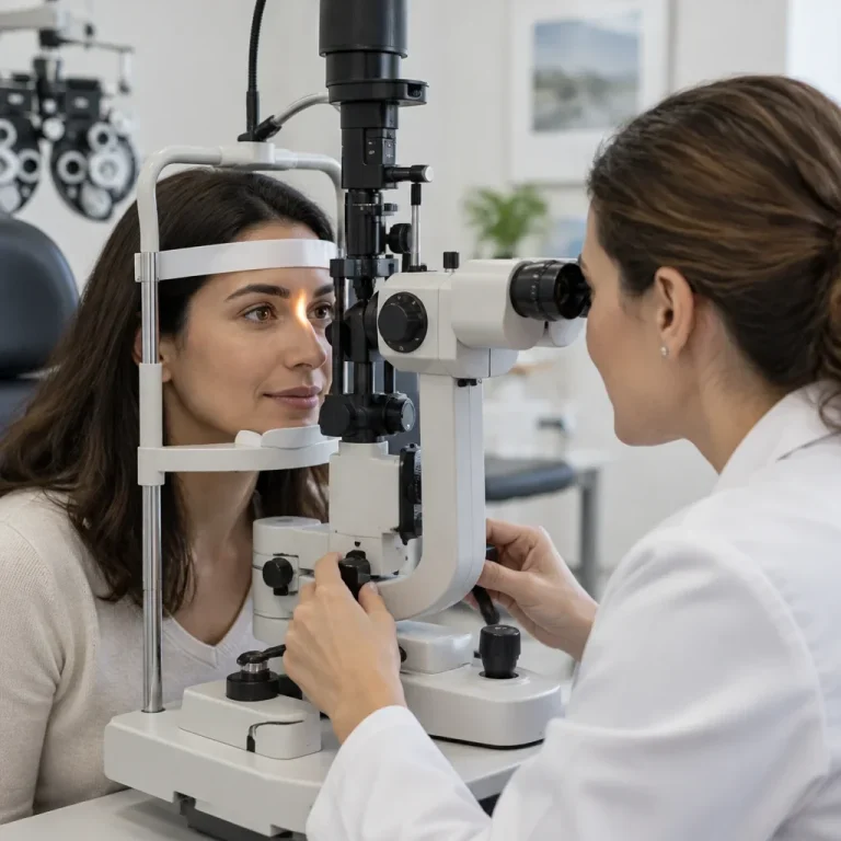
Clear vision does not mean diabetes has left your eyes unaffected. Diabetes and eye tests belong together because retinal damage can develop without noticeable symptoms. A diabetic eye exam looks for that damage, but it cannot diagnose or rule out diabetes by itself.
Diabetes and eye tests: more than a prescription check
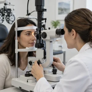
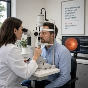
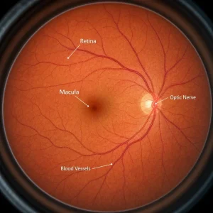
A diabetic eye exam assesses eye health, particularly the retinal blood vessels that diabetes can damage. The retina is the light-sensitive tissue lining the back of your eye. Checking how clearly you read an eye chart does not establish whether those vessels are healthy.
A comprehensive examination can include both prescription testing and retinal assessment. The difference is not simply “regular” versus “diabetic.” Your diabetes history, the retinal examination and the follow-up plan determine whether the assessment addresses diabetes-related risks.
| Assessment | Main purpose | Important limitation |
|---|---|---|
| Prescription-only vision testing | Measures focusing and the correction needed for clearer vision | Does not establish retinal health |
| Comprehensive eye examination | Assesses vision and eye health, including structures at the front and back of the eye | Additional imaging or specialist assessment may still be needed |
| Diabetes-focused retinal screening | Looks for diabetes-related retinal changes, sometimes using photographs | Screening alone may not assess your prescription, cataracts, glaucoma or the entire eye |
A DM eye exam means an examination related to diabetes mellitus. “Diabetic vision exam,” “diabetic fundus exam” and “retinopathy screening” are related descriptions, but their scope can differ. Tell the optometrist you have diabetes rather than assuming every vision test includes the same retinal assessment.
Can an eye test detect diabetes?
An eye examination can reveal findings that raise suspicion of diabetes, but blood testing establishes the diagnosis. Blood glucose and A1C testing assess different aspects of blood sugar. A normal retinal examination does not prove that either is normal.
Retinal bleeding, leaking vessels and fluctuating focus can prompt further investigation. These findings are not specific proof of diabetes: high blood pressure and other conditions can cause overlapping changes. Detecting possible diabetes during an eye exam means identifying a reason for medical assessment, not making a diagnosis from a retinal image alone.
A “glucose eye test” is not a replacement for blood glucose or A1C testing. Changes in blood sugar can alter the eye’s focusing tissues and temporarily change vision, but an ordinary eye examination does not measure your blood glucose level.
An optometrist examines eye health and can identify suspected diabetic eye disease. An optician fits and dispenses glasses and contact lenses; that role does not include diagnosing diabetes. Physicians and nurse practitioners assess diabetes using medical history and appropriate laboratory testing.
Eye symptoms that need prompt or emergency care
Early diabetic retinopathy can cause no noticeable symptoms. There is no dependable pair of “first symptoms” that makes waiting safe. Blur and floaters can occur, but their absence does not remove the need for screening.
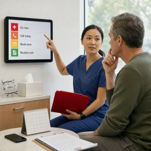
Urgent warning signs need same-day assessment, not routine online booking:
- Sudden loss of vision or a sudden major reduction in vision.
- A curtain, shadow or missing area in your vision.
- New flashes accompanied by a sudden increase in floaters.
- Severe eye pain, particularly with redness, halos, headache or nausea, including after dilation.
Go to an emergency department if timely urgent eye care is unavailable. These symptoms can indicate retinal detachment, bleeding or another sight-threatening condition, whether or not diabetes is the cause.
New fluctuating blur, difficulty reading or gradual changes in vision also need an appointment. An examination can distinguish a prescription change from cataract, retinal swelling or another eye problem. Do not assume that every change is simply your blood sugar settling.
When people with diabetes should have eye exams
Screening begins according to diabetes type, then follows your retinal findings and individual risk. Diabetes duration, glucose management, blood pressure, pregnancy and previous eye disease all influence the next appointment. Clinical follow-up and insurance eligibility are separate decisions.
| Situation | Initial assessment or follow-up approach |
|---|---|
| Newly diagnosed adult with type 2 diabetes | Retinal assessment at diagnosis; diabetes may have been present before it was recognized. |
| Adult with type 1 diabetes | Initial retinal assessment within 5 years of diagnosis, followed by regular examinations. |
| Adult with type 1 diabetes and normal retinal findings | Annual screening is the usual starting schedule; the clinician adjusts follow-up to findings and risk. |
| Type 2 diabetes with no retinopathy | A 1–2-year interval can be appropriate when the clinician considers the findings and risk factors suitable. |
| Existing retinopathy, suspected swelling or changing findings | Follow-up is individualized and may be more frequent than routine screening. |
| Pre-existing diabetes when planning pregnancy | Arrange assessment before conception when possible, with early pregnancy assessment and subsequent monitoring directed by findings. |
| Gestational diabetes alone | Does not automatically require the retinal screening schedule used for pre-existing diabetes; symptoms or other concerns still require assessment. |
Children and adolescents need age- and diabetes-duration-specific screening plans. The adult type 1 schedule should not simply be applied to every child. Pregnancy also requires an individualized plan, including follow-up after delivery where indicated.
A normal result means no retinopathy was detected at that assessment, not that future damage is impossible. Risk increases with diabetes duration, but there is no dependable number of symptom-free years during which screening can be skipped.
What happens during a diabetic eye exam
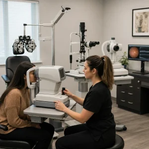
A diabetes-focused examination combines your medical history with vision and eye-health assessment. The exact tests depend on the findings, your previous records and whether the retina can be examined adequately.
- Review your health history. The optometrist asks about diabetes type, diagnosis date, medications, blood pressure, pregnancy and previous retinal treatment. Bring your recent A1C result if available.
- Assess vision and focusing. Reading charts and prescription testing help identify reduced vision and whether focusing changes contribute.
- Examine eye structures and pressure. Assessment can identify cataracts, pressure-related concerns and other conditions alongside diabetic disease.
- Examine the retina. Dilating drops, retinal imaging or a combination may be used to assess blood vessels, the macula and the optic nerve.
- Discuss the findings and next steps. The plan may involve routine monitoring, additional testing or referral for specialist assessment.
Dilation enlarges the pupil so the clinician can obtain a better view inside the eye. The drops do not measure blood sugar. A fundus examination is generally not painful, although drops can briefly sting and the examination lights can feel bright.
The appointment includes more than the examination itself when dilation is needed. Drops require time to take effect, and additional imaging or assessment can extend the visit. When arranging an appointment at Premier Optical, mention diabetes and ask how much time to allow for the planned examination.
Findings visible during the examination can be explained at the visit. A specialist consultation or additional testing may be needed before there is a complete diagnosis or treatment plan. Clarify which results are available and how any outstanding findings will be communicated.
What the different retinal tests show
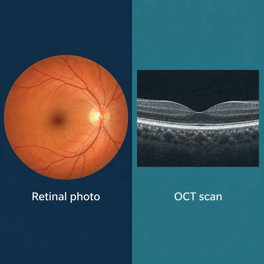
Retinal tests answer different clinical questions and are not interchangeable. A photograph records appearance; a cross-sectional scan can show swelling within retinal layers. Neither automatically replaces a comprehensive examination.
| Test | What it assesses | What you experience | When it may be used |
|---|---|---|---|
| Fundoscopy or ophthalmoscopy | The retina, retinal blood vessels and optic nerve | Bright lights and examination through a viewing instrument, sometimes after dilation | Direct assessment of the back of the eye |
| Fundus photography | A recorded image of the retinal surface | A camera image, sometimes with a bright flash | Screening, documentation and comparison over time |
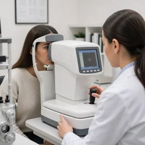
| Optical coherence tomography, or OCT | Cross-sectional views of retinal tissue, including the macula | Looking into a scanner; no injection is needed for the scan | Assessing suspected macular swelling or other structural changes |
| Tonometry | Pressure inside the eye | The technique may use a gentle probe or air puff | Assessing eye pressure; it does not diagnose retinopathy by itself |
| Fluorescein angiography | Retinal circulation and leakage | Dye is injected into a vein, followed by retinal photographs | Specialist investigation when the findings require it, not routine testing for everyone |
Non-dilated photographs can be useful in selected screening settings, but they are not an automatic substitute for dilation. Small pupils, cataracts, image quality and the area captured can limit what is visible. An inadequate image needs further assessment rather than a reassuring “normal” label.
Preparing for the exam and getting home safely
Routine diabetic eye assessment generally does not require fasting. Keep your usual meal and medication routine unless you have specific medical instructions for another procedure. Do not change diabetes medication simply because an eye examination is booked.
Bring the information and items that help the examination proceed:
- Your current glasses and contact lens information.
- Your medication list and recent A1C result, if available.
- Your Ontario health card and private insurance details.
- Previous eye reports, if you already have them.
- Details of your diabetes care provider and previous retinal treatment.
- Sunglasses for the trip home after dilation.
Dilating drops can cause blurred near vision and light sensitivity for approximately 2–6 hours, with effects sometimes lasting longer depending on the drops and your response. Arrange transportation when dilation is expected, particularly if you have not experienced it before.
After dilation:
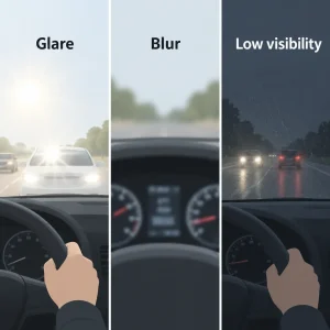
- Do not drive or operate machinery while blur or glare affects safe vision.
- Use sunglasses if bright light is uncomfortable.
- Allow for temporary difficulty with reading and screens.
- Do not use an elapsed number of hours as automatic clearance to drive.
- Seek urgent assessment for severe pain, marked redness, nausea or sudden worsening vision.
Mobility, communication and support-person needs can affect how an examination is arranged. If you need help transferring, positioning at equipment or understanding instructions, tell Premier Optical when booking so the team can discuss appointment arrangements with you.
What diabetic eye exam results mean
Diabetic retinopathy is damage to retinal blood vessels associated with diabetes. Diabetic macular edema is fluid-related swelling in the macula, the area responsible for detailed central vision. Macular edema can occur at different retinopathy stages.
| Result or finding | Plain-English meaning | General next step |
|---|---|---|
| No retinopathy detected | No visible diabetic retinal damage was identified in an adequate assessment | Continue screening and diabetes management; this does not rule out diabetes |
| Non-proliferative retinopathy | Retinal vessels show changes such as small bulges, bleeding or leakage, without abnormal new vessel growth | Monitoring and referral depend on severity and associated findings |
| Proliferative retinopathy | Abnormal new vessels have developed; these can bleed and contribute to scar tissue | Specialist assessment and treatment planning |
| Suspected macular edema | Fluid may be affecting the central retina | Further assessment, often including OCT and specialist review |
| Inadequate or unclear images | The retina could not be assessed reliably from the images | Repeat imaging, dilation or another examination; not a normal screening result |
“Background retinopathy” usually describes earlier non-proliferative changes. “Referable retinopathy” means the findings meet that screening service’s criteria for further assessment. These terms do not replace an explanation of your actual findings or establish a universal Ontario treatment category.
Before leaving, ask for a clear explanation of the result and follow-up plan:
- Was retinopathy or macular swelling found?
- Has anything changed since the previous examination?
- When is the next examination needed?
- Is specialist referral necessary?
- Will a report be shared with the diabetes care team?
Treatment, prevention and the limits of reversal
Some early retinal changes can improve with appropriate management, but established damage is not reliably reversible. Lost vision may not return. “Stage 1” is not a promise that the condition can be cured, and treatment depends on the actual retinal findings rather than a stage label alone.
Specialist treatment can include anti-VEGF injections to reduce leakage or abnormal vessel activity, retinal laser and vitrectomy surgery. Vitrectomy removes the vitreous gel and can address bleeding or traction in selected cases. Not every person with retinopathy needs an injection, laser or surgery.
Sustained high glucose, high blood pressure, kidney disease, diabetes duration and pregnancy can influence progression. Individualized glucose, blood pressure and lipid management, smoking cessation support and regular retinal assessment all form part of diabetes eye care. These are coordinated medical decisions, not reasons to adjust medication independently.
Blindness is not inevitable with diabetic retinopathy. Monitoring and indicated treatment can help protect vision, but neither a normal result nor treatment guarantees that disease will never progress. Keeping follow-up appointments matters even when you see clearly.
Blurred vision from changing glucose levels differs from blur caused by macular edema, cataracts or glaucoma. Diabetes increases cataract risk and is associated with increased glaucoma risk. Glasses can correct focusing errors, but they do not treat retinal bleeding, swelling or nerve damage.
A fluctuating prescription may need reassessment once glucose becomes more stable. There is no universal duration for diabetes-related blur and no single A1C level at which eye damage starts. New or sudden blur needs assessment rather than simply waiting for blood sugar to improve.
OHIP and private insurance coverage in Ontario
OHIP-insured people with diabetes are eligible for a major eye examination every 12 months under Ontario’s diabetes-related eye-care coverage. That does not mean every imaging test or additional service is automatically insured.
An insured major examination, medically necessary follow-up assessment and an uninsured imaging service are different billing categories. Retinal photography and OCT should not be assumed to be included or excluded solely because diabetes is present. The service, its clinical purpose and the applicable coverage rules determine billing.
Clinical follow-up can be needed sooner than the interval for another insured major examination. Payment eligibility should not be used as a reason to delay assessment of new symptoms or abnormal findings.
Private benefits may contribute to eligible uninsured examinations or imaging, according to the plan’s terms. Premier Optical offers direct insurance billing, but direct billing does not guarantee reimbursement. Deductibles, benefit limits and eligible services determine any remaining balance.
Accessing diabetic eye care in Waterloo and Kitchener
An optometrist is an appropriate starting point for comprehensive diabetic eye assessment and ongoing monitoring. Ophthalmologists are medical and surgical eye specialists; a retina specialist manages retinal disease when specialist investigation or treatment is indicated.
- Office-based optometry: Comprehensive examination, detection, monitoring and referral when needed.
- Ophthalmology or retinal care: Specialist assessment and medical or surgical treatment according to the condition.
- Community or teleophthalmology screening: Retinal images are captured and interpreted through a screening pathway. This does not necessarily replace comprehensive eye care, and an abnormal or unclear result needs follow-up.
A routine optometrist appointment can generally be booked directly without a physician referral. Specialist appointments and particular screening pathways can have different referral requirements. Retinal care may also involve sharing examination reports with your medical team, with appropriate consent.
Premier Optical brings optometrist examinations and optical services together in Waterloo. That coordination can help with vision assessment and prescription needs, but glasses and frame fitting do not treat diabetic retinal disease. Specialist referral remains necessary when the findings call for it.
Frequently asked questions about diabetes and eye tests
A regular eye exam and suspected diabetes
A comprehensive eye examination can uncover changes suggestive of diabetes if it includes retinal assessment. A prescription check alone cannot reliably do that. Neither examination establishes or excludes diabetes without the appropriate medical assessment and blood testing.
Screening when glucose is well controlled
Retinal screening remains necessary even when your current glucose results are within your agreed targets. Eye health reflects more than a recent blood result, including diabetes duration, previous glucose exposure, blood pressure and existing retinal changes.
Retinopathy without diabetes
Retinopathy can occur without diabetes. High blood pressure, retinal vein occlusion and other vascular conditions can cause bleeding or changes that overlap with diabetic findings. Diagnosis requires the pattern of findings, examination and medical history, not comparison with an online image.
Diabetes-related blur and a new glasses prescription
A new prescription may improve a focusing problem, but the cause of blur must be assessed first. Glucose fluctuations can change focusing, while retinal swelling or cataracts need a different management plan. There is no fixed waiting period that makes unexplained blur safe to ignore.
An eye examination without dilating drops
Some retinal screening can be performed without dilation, but the clinician must be able to obtain an adequate assessment. Image quality, pupil size, symptoms and previous findings help determine whether drops or further examination are needed.
For routine care, book an eye exam at Premier Optical in Waterloo and mention diabetes when scheduling. Bring your medication information and previous eye-care details. The useful outcome is more than a prescription: it is a clear explanation of your retinal health and your next step.