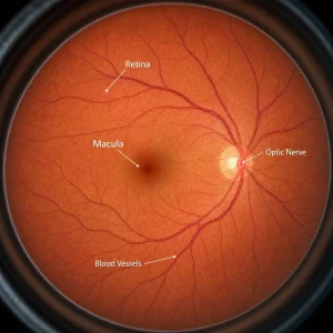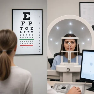
An abnormal OCT eye test does not automatically mean a severe eye disease or future blindness. It means the scan showed a structural change that an optometrist or ophthalmologist needs to interpret with the rest of your exam.
What an abnormal OCT eye test means
An OCT eye exam creates cross-section images of the retina and optic nerve at the back of the eye. OCT stands for optical coherence tomography, and it shows layers of tissue rather than just a surface photo .
An abnormal result usually means the scan found something that looks different from the expected structure for that part of the eye. That can include fluid, swelling, thinning, deposits, traction, a break in normal layers, or loss of nerve tissue .
An abnormal OCT scan is not a diagnosis on its own. OCT eye test results are one part of a full assessment that may also include your symptoms, vision testing, dilated examination, retinal photos, pressure testing, and sometimes visual field testing .
Some OCT findings are mild and only need monitoring over time. Others need prompt referral, and sudden symptoms can make the same finding much more urgent .
This page explains what common patterns can mean in plain language. It cannot diagnose your scan online, and a clinician still needs to review the image in context .
Why an optometrist may recommend an OCT eye exam
An optometrist may recommend an OCT eye exam when your symptoms suggest a structural problem that cannot be fully judged from symptoms alone. Common reasons include blurred central vision, distortion, flashes, floaters, diabetes, glaucoma risk, macular degeneration risk, unexplained vision changes, or a need for baseline monitoring .
An OCT retina scan can reveal changes that may not be obvious early on. That matters because some retinal and optic nerve diseases begin with tissue changes before the patient notices major vision loss .
Follow-up is another common reason for OCT imaging. If a prior scan showed fluid, drusen, nerve fiber thinning, or traction, repeat scans help compare whether the structure is stable, improving, or worsening over time .
New visual symptoms should lead to a comprehensive eye exam rather than waiting to see if they pass. Sudden distortion, a dark curtain, or a rapid change in one eye should not be watched at home .
Normal vs abnormal OCT: what doctors look for
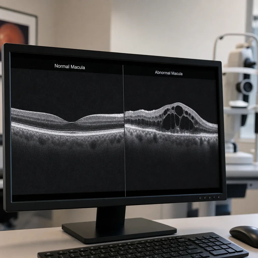
A normal macular scan usually shows a smooth retinal contour, an expected foveal dip in the centre, organized retinal layers, and no fluid pockets or suspicious elevations. That is the basic pattern behind normal vs abnormal OCT eye test comparisons .
A normal optic nerve OCT usually shows nerve fiber and related layer thickness within the expected range for that patient, with no suspicious pattern of thinning or asymmetry that fits disease. The optic nerve assessment depends on more than one scan and must be matched with the rest of the exam .
An abnormal scan often shows one or more structural changes. Common examples are retinal thickening, thinning, cystic spaces, subretinal fluid, intraretinal fluid, drusen, epiretinal membrane, traction, or thinning in the retinal nerve fiber layer around the optic nerve .
A normal OCT scan does not always rule out disease, and an abnormal one does not always confirm it. Image quality, anatomy, age, previous disease, and correlation with symptoms all affect interpretation .
| OCT finding | What it may suggest | Common symptoms | Usual next step |
|---|---|---|---|
| Intraretinal fluid | Swelling in the retina, sometimes from diabetic macular edema or wet macular disease | Blurred or distorted central vision | Prompt exam review or referral |
| Subretinal fluid | Fluid under the retina, seen in some macular disorders | Distortion, blur, central patchiness | Prompt review |
| Drusen | Deposits linked with age-related macular degeneration | Often none early, later central vision change | Monitor or refer based on severity |
| Epiretinal membrane | Thin scar-like tissue pulling on the retina | Wavy lines, distortion | Monitor or refer if symptomatic |
| Macular hole or lamellar hole | Structural defect in the macula | Distorted or missing central spot | Referral depending on stage |
| RNFL thinning | Possible glaucoma or other optic nerve damage | Often no early symptoms | Correlate with pressure, fields, and exam |
| Poor-quality scan or artifact | Image may be unclear or misleading | Symptoms may not match | Repeat scan or different testing |
How to read common terms on an OCT report
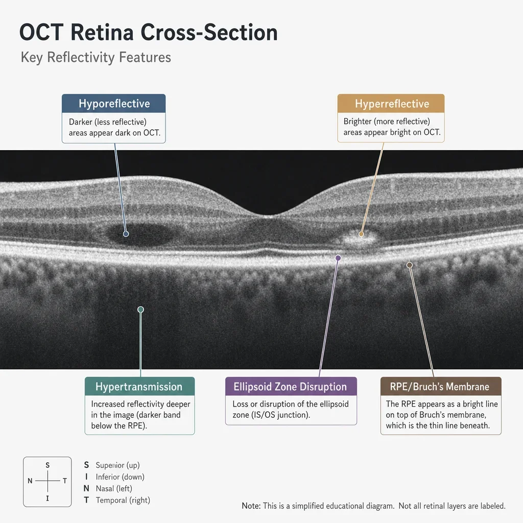
Hyporeflective means a darker area on the scan. Depending on where it sits, it may represent a normal low-reflective space or fluid within or under the retina .
Hyperreflective means a brighter area on the scan. Depending on location, it may reflect a normal bright layer, a membrane, deposits, scarring, or inflammatory material .
Hypertransmission means more light is passing into deeper tissue than expected. That can happen when tissue above is thinned or lost, which is why the term is often discussed in macular disease and atrophy .
Ellipsoid zone disruption means a change in an outer retinal band linked to photoreceptor health. When that band is disrupted, visual quality may be reduced even if the rest of the scan looks less dramatic .
RPE means retinal pigment epithelium, and Bruch’s membrane is a thin support layer beneath it. Changes or disruption there can be part of age-related macular degeneration and other retinal disease patterns .
The impression section on a report matters more than any single word. A technical term without the clinician’s interpretation is not enough to tell you what condition is present or how serious it is .
What diseases an OCT scan can detect or help monitor
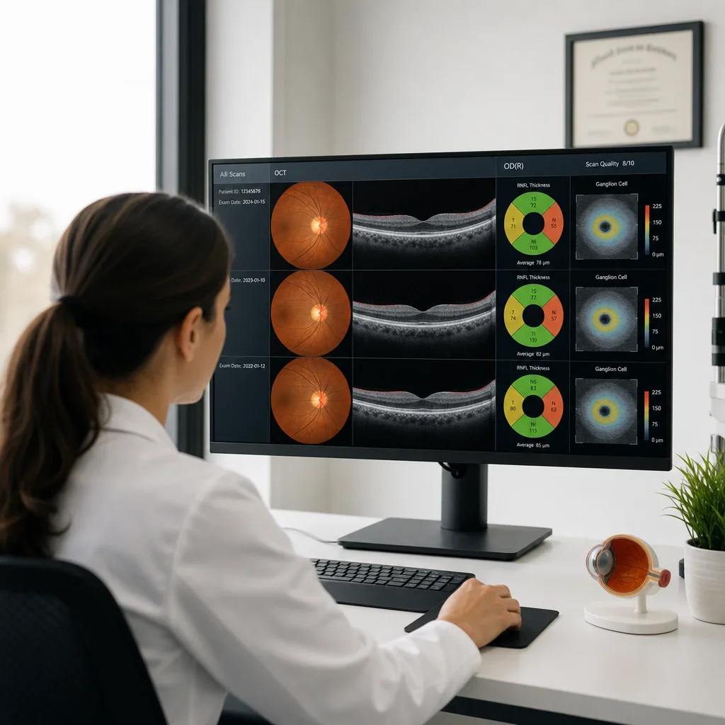
OCT can help detect or monitor disease in the retina, macula, vitreomacular interface, and optic nerve. It is especially useful for structural problems that change retinal thickness, retinal layers, or nerve tissue appearance .
Common retinal OCT abnormalities include age-related macular degeneration, diabetic macular edema, epiretinal membrane, vitreomacular traction, macular hole, and some vascular or inflammatory causes of swelling. These patterns can overlap, so final diagnosis still depends on the full eye exam .
Common optic nerve OCT abnormalities include glaucomatous thinning and changes that can appear after optic neuritis or other optic neuropathies. OCT supports assessment, but it does not replace pressure measurement, nerve examination, and visual field testing .
OCT is a strong monitoring tool because the same area can be scanned again and compared over time. That makes it useful for glaucoma, diabetic eye disease, macular degeneration, and previously documented retinal changes .
Retinal diseases commonly linked to an abnormal OCT eye test
Age-related macular degeneration is one of the most common reasons for an abnormal macular scan. In dry disease, the OCT may show drusen, pigment epithelial changes, or areas of thinning and atrophy, while wet disease may show intraretinal or subretinal fluid and sometimes elevation from abnormal blood vessel growth .
Drusen usually appear as bumps or elevations under the retina at the level of the retinal pigment epithelium. They can be small and stable, or they can be larger and associated with more significant AMD changes over time .
In dry AMD or geographic atrophy, the scan may show thinning and loss of outer retinal layers, plus increased signal into deeper tissue that is described as hypertransmission. In wet AMD, fluid is the key finding that raises concern for active leakage .
OCT for diabetic retinopathy is especially useful when fluid affects the macula. Diabetic macular edema can appear as retinal thickening, cystic spaces, or fluid under the retina, and serial scans help show whether swelling is stable or changing .
An epiretinal membrane appears as a thin layer on the retinal surface that can wrinkle or distort the macula. Patients may notice wavy lines, smaller-looking images, or blurry central vision when the traction is significant .
Vitreomacular traction happens when the vitreous pulls on the macula instead of separating cleanly. That traction can distort the retinal contour and may lead to symptoms even before a full macular hole forms .
A lamellar hole is a partial-thickness defect, while a full-thickness macular hole extends through the retinal layers at the macula. Both can affect central vision, but their OCT appearance and urgency differ .
Other retinal OCT abnormalities can be seen with central serous-type fluid patterns, retinal vein-related swelling, inflammatory disease, and some inherited retinal disorders. OCT helps show structure, but the cause still needs clinical interpretation .
Optic nerve abnormalities OCT may show
OCT for glaucoma looks at the retinal nerve fiber layer and related structures around the optic nerve. Thinning in characteristic patterns can support concern for glaucoma, but diagnosis also depends on eye pressure, optic nerve appearance, corneal factors, and visual field testing .
The first signs of optic nerve damage are not always symptoms. Early glaucoma may cause no noticeable vision change, which is why structural imaging and routine exams matter for at-risk patients .
When symptoms do appear, optic nerve problems can cause reduced side vision, blur, lower contrast, washed-out colours, or trouble in dim light. Those symptoms are not unique to one disease, so they still need examination .
Two diseases that can affect the optic nerve are glaucoma and optic neuritis. Other optic neuropathies also exist, but these two are among the best-known examples patients hear about in clinic .
Optic nerve OCT abnormalities can also appear after inflammation or damage to the nerve pathway. OCT can show thinning over time after optic neuritis, but it does not diagnose multiple sclerosis by itself .
Symptoms that can match abnormal OCT findings
Distorted central vision can match macular swelling, traction, or a membrane affecting the macula. Wavy lines are a classic symptom of macular distortion, but the cause can range from fluid to scar-like surface traction .
Blurred or patchy central vision can line up with fluid, atrophy, or a developing hole in the macula. Reduced reading clarity is a common complaint when the centre of the retina is affected .
Flashes and floaters can be harmless, but sudden new flashes with many floaters can signal a retinal tear or detachment. Those symptoms are urgent even before OCT results are discussed .
A curtain, shadow, or missing part of the side vision is a red flag in the eye. That symptom can point to retinal detachment and needs same-day urgent assessment .
Symptoms and scan findings do not match one-to-one. Some people have clear OCT changes with few symptoms, and others have symptoms that need different testing even if the OCT is less dramatic .
Urgent warning signs: when an abnormal OCT or eye symptoms need same-day care

Sudden vision loss, a new shower of floaters, repeated flashes, a curtain or shadow, severe new distortion, pain with vision loss, or sudden dimming in one eye should be treated as urgent. These are red flag eye symptoms that need same-day assessment .
The early warning signs of a detached retina often include flashes, floaters, and a dark curtain or veil in the vision. Retinal detachment is a medical urgency, and waiting for a routine appointment can risk permanent vision loss .
Some abnormal OCT findings are not emergencies. Stable drusen, mild membrane changes, or monitored thinning may only need scheduled follow-up if symptoms are unchanged .
Sudden symptoms change the urgency even when the scan finding sounds familiar. A patient with known macular disease who develops rapid new distortion or vision loss needs faster review than a patient with a stable chronic finding .
> Urgent red flags callout > Same-day urgent assessment: sudden vision loss, curtain or shadow, new flashes with many floaters, pain with vision loss, sudden one-eye dimming > Book promptly: new distortion, new central blur, worsening reading vision, a recent abnormal scan without clear follow-up > Routine follow-up: stable findings already being monitored, no new symptoms, repeat imaging planned by your clinician
What to do after an abnormal OCT result
The next step after an abnormal result is to have the scan reviewed with a full eye exam if that has not already happened. That may include dilation, retinal photos, visual field testing, repeat OCT imaging, or referral to an ophthalmologist depending on the pattern and your symptoms .
Ask for the practical summary, not just the technical wording. The most useful questions are what was seen, whether it is urgent, whether you need dilation, whether referral is needed, when to return, and what symptoms should make you come back sooner .
Repeat imaging is common when the first scan is borderline, the image quality is poor, or the clinician wants to compare a baseline with a later scan. Monitoring over time is one of the main strengths of OCT eye test results .
Do not try to judge seriousness from one term on a portal or printout. A word like fluid, thinning, or disruption needs location, severity, and exam correlation before it means anything useful for your own eye health .
If you were told you had an abnormal OCT elsewhere and still do not understand the plan, booking a comprehensive eye exam in Waterloo for imaging review is a reasonable next step. The goal is clarification, not self-diagnosis .
Can an abnormal OCT mean blindness or permanent damage?
An abnormal OCT does not automatically mean blindness. It means there is a structural finding that needs to be explained and managed according to its cause, location, and severity .
Some causes are treatable or manageable, especially when found early. Fluid-related changes, traction problems, and some inflammatory causes may improve with the right care, while other structural damage may be long-standing or permanent .
Permanent damage is more concerning when there is clear tissue loss, advanced atrophy, or long-standing optic nerve loss. Even then, the purpose of diagnosis and monitoring is to protect remaining vision and guide treatment decisions .
No online guide can tell you whether a change is reversible from the image wording alone. Prognosis depends on the disease, how early it was found, and how quickly appropriate care happens .
Can OCT detect MS or other non-retinal problems?
OCT can show changes related to optic nerve damage or retinal nerve fiber loss in some patients with optic neuritis and some neurologic disease. It can support the picture, but it cannot diagnose multiple sclerosis on its own .
First eye-related signs that can happen with optic neuritis include pain on eye movement, blurred vision in one eye, washed-out colour vision, and reduced contrast. Those symptoms warrant prompt medical evaluation .
If a clinician suspects a neurologic cause, the next step is referral and broader medical workup rather than relying on OCT alone. OCT is one tool in a larger assessment pathway .
Limitations of OCT: artifacts, unclear scans, and why repeat imaging happens
OCT can be wrong or unclear when the scan quality is poor. Blinking, dry eyes, poor fixation, cataract or other media opacity, shadowing, and segmentation errors can all affect the image and make interpretation less reliable .
A segmentation error means the machine placed its measurement lines on the wrong layers. That can create misleading thickness maps or false-looking abnormalities unless a clinician notices the mistake .
A repeat OCT scan may be done to get better alignment, confirm a suspicious area, or compare a finding over time. One imperfect scan does not necessarily mean disease .
This is one reason patients should not self-interpret colour maps or thickness printouts. The image itself, the signal quality, and the exam findings all matter together .
What the OCT test is like: pain, dilation, time, and repeat visits
An OCT scan is generally non-contact and painless. You usually place your chin on a support and look at a target while the machine captures the image .
The scan itself usually takes only a few minutes , while the full visit length varies with the reason for the exam, whether dilation is needed, and whether other testing is done.
Dilation is not always required for OCT, but some visits still need it. If the clinician needs a wider retinal view, clearer assessment of the back of the eye, or a fuller examination because of symptoms or disease, dilating drops may be recommended .
Repeat scans are common for glaucoma, macular degeneration, diabetic eye disease, and any prior abnormality being followed over time. The point is to compare structure from one visit to the next, not just to take another picture .
FAQ about abnormal OCT eye test results
What does an abnormal OCT eye test mean?
It means the scan showed a structural change in the retina or optic nerve that needs clinical interpretation. It is not a diagnosis by itself .
What diseases can an OCT scan detect?
OCT can help detect or monitor glaucoma, macular degeneration, diabetic macular edema, epiretinal membrane, vitreomacular traction, macular hole, and other retinal or optic nerve disorders .
Why would a doctor recommend an OCT eye exam?
Common reasons include blurred or distorted vision, flashes or floaters, diabetes, glaucoma risk, macular degeneration risk, unexplained vision changes, or follow-up of a known finding .
Is an abnormal OCT serious?
It can be mild, moderate, or urgent depending on the pattern and your symptoms. Sudden symptoms always raise the urgency .
Does an abnormal OCT mean blindness?
No. An abnormal result does not automatically mean blindness, but it does mean you need proper assessment and follow-up .
Can an abnormal OCT be reversed?
Some causes can improve or be managed, while other structural damage may be permanent. The cause matters more than the word abnormal itself .
What are the first signs of retinal disease?
Common early signs include distortion, wavy lines, blurred central vision, patchy missing areas, new flashes, or new floaters. Some retinal disease causes few symptoms at first .
What are the first signs of optic nerve damage?
Early optic nerve damage may cause no symptoms. Later signs can include reduced side vision, dimmer vision, and changes in contrast or colour perception .
What are the early warning signs of a detached retina?
New flashes, a sudden increase in floaters, and a curtain or shadow in the vision are classic warning signs and need same-day assessment .
Can OCT detect glaucoma?
OCT can detect structural thinning that may support glaucoma assessment and monitoring, but it does not diagnose glaucoma by itself .
Can OCT detect macular degeneration?
OCT is very useful for showing drusen, fluid, atrophy, and other structural changes linked with macular degeneration .
Can OCT detect diabetic eye disease?
OCT is useful for diabetic macular edema and other retinal swelling, especially when the macula is involved .
Can OCT detect MS?
OCT cannot diagnose multiple sclerosis on its own. It may show nerve fiber loss related to optic neuritis or other neurologic disease, but diagnosis requires broader medical assessment .
Do I need dilation for an OCT eye exam?
Not always. Some OCT scans are done without dilation, but dilation may still be recommended based on symptoms, exam findings, or the need for a better retinal view .
Is an OCT eye exam painful?
No. OCT is typically painless and non-contact .
How long does an OCT eye exam take?
The scan itself usually takes a few minutes , but the full visit can take longer if you need dilation or other testing.
What should I do after an abnormal OCT result?
Make sure a clinician reviews the scan with the rest of your exam, ask whether it is urgent, and follow the recommended plan for repeat imaging, monitoring, or referral .
When to book an eye exam in Waterloo
Book an eye exam if you were told you had an abnormal OCT scan, if you have new visual symptoms, or if you are due for monitoring of glaucoma, diabetes, or macular disease. A full assessment matters more than trying to decode the report yourself .
If you need an eye exam Waterloo patients can use for clarification after abnormal imaging, the practical next step is a comprehensive visit with an optometrist who can review symptoms, examine the back of the eye, and explain whether monitoring or referral is needed .
For local patients looking for an optometrist Waterloo eye clinic, Premier Optical offers comprehensive eye exams, optician support, and streamlined care under one roof. If you have questions about an abnormal result or need baseline monitoring, booking an exam is the safest next step.
If your symptoms are sudden or severe, do not wait for a routine appointment. Seek same-day urgent assessment instead .