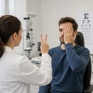
A 60 4 visual field test measures side vision, not how clearly you read letters. That confusion trips people up all the time. This guide explains what a 60 4 visual field is, how the test works, when clinicians choose it, and what a Humphrey visual field report may actually show.
What the 60 4 visual field test is
The 60 4 visual field test is a wide-field automated perimetry pattern used to check more peripheral vision than standard central field tests such as 24-2 or 30-2. Visual field testing maps how well you detect lights in different directions while you keep looking straight ahead, and the goal is to find areas where sensitivity is reduced or missing.
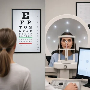
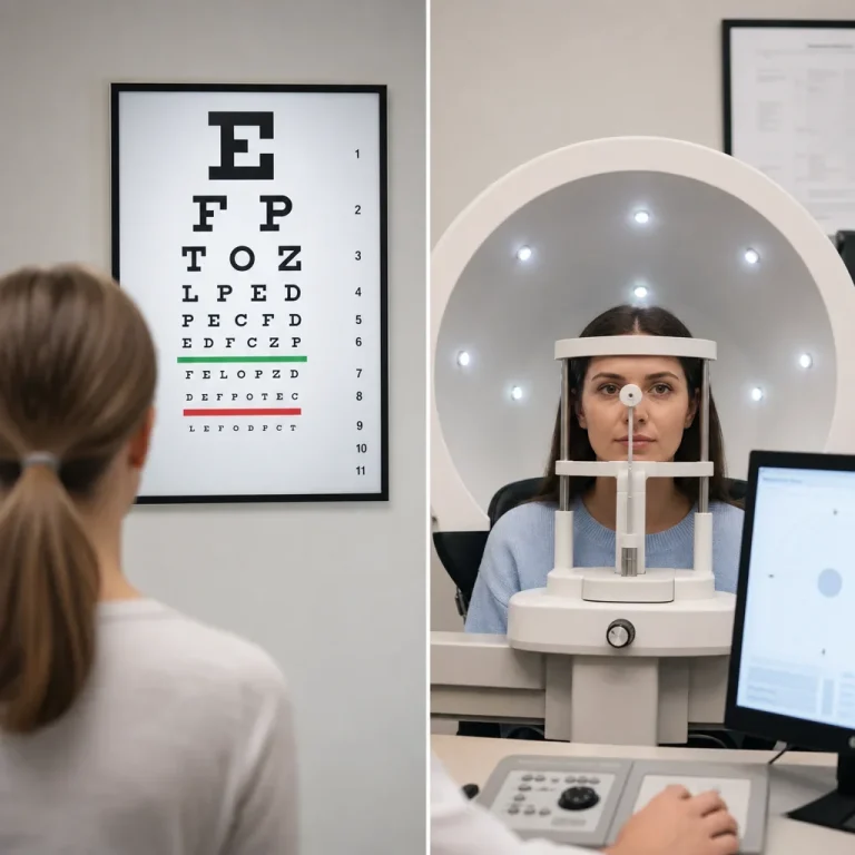
A 60/4 visual field is not the same as visual acuity. Visual acuity is the chart reading number like 20/20, 20/40, or 6/60, while a visual field test measures how much of the space around you you can detect without moving your eyes.
The naming of visual field programs refers to the test pattern and area being sampled, but exact point layouts can vary by machine model and software version. That is why we tell patients to treat the program name as a clue, not the whole story, and to ask the clinic what device and strategy were used. This is general information, not an interpretation of your own results.
What the test measures and what it does not
A visual field test measures where you can see around you, not how sharp the image is. Someone can have excellent central acuity on a chart and still have major peripheral loss, or the reverse can happen.
A 60 degree field of view and a 70 degree field of vision are not the same thing as 6/60 eyesight. The first terms describe spatial extent, while 6/60 is an acuity measurement that means a person sees at 6 metres what a person with standard acuity sees at 60 metres.
Legal blindness and driving questions cannot be answered from a 60-4 result alone. In practice, those decisions may involve acuity, visual field, medical review, and the rules of the jurisdiction, so patients should use official Ontario or Canadian sources and their eye care provider rather than internet shortcuts.
How a 60 4 visual field test works during the appointment
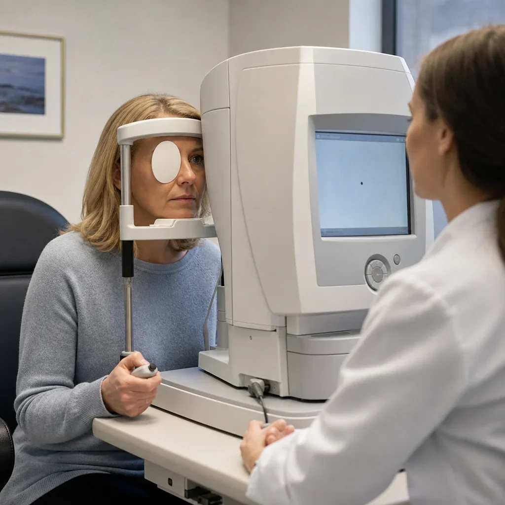
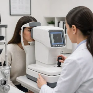
The Humphrey visual field test procedure is straightforward: one eye is tested at a time while you rest your chin and forehead on the machine and press a button whenever you see a light. The other eye is covered, and you are asked to keep looking at the central target instead of chasing the flashes.
Most automated visual field tests take about 5 to 15 minutes per eye depending on the program, patient reliability, pauses, and whether any part needs to be repeated. A wider or more demanding test can feel longer because peripheral testing may require more attention and steadier positioning.
Blinking is allowed and usually helps comfort. Dry eyes, fatigue, misunderstanding the instructions, or poor positioning can all reduce reliability, so a technician may stop to coach you, reposition you, or restart part of the test.
Repeat testing is common when the first run looks unreliable or does not fit the symptoms and exam. That is not a failure. It is part of getting a result worth trusting.
60-4 vs 60-2 vs 24-2 vs 30-2
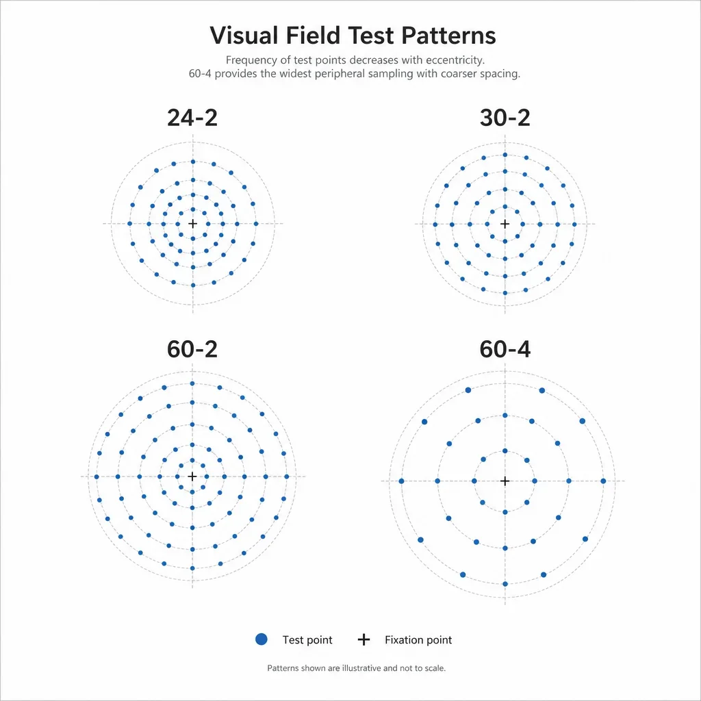
The main difference between 60-4, 60-2, 24-2, and 30-2 visual field tests is the area sampled and the spacing of the test points. Central programs such as Humphrey visual field 24-2 and the 30 2 visual field test are commonly used when the main question is central or mid-peripheral loss, while 60-series patterns are chosen when the clinician wants a wider look into the peripheral field.
| Test pattern | Area tested | Point spacing | Common use | Main limitation |
|---|---|---|---|---|
| 24-2 | Central field | Moderately spaced central grid | Routine glaucoma assessment and follow-up | Misses far peripheral loss outside the central area |
| 30-2 | Slightly wider central field than 24-2 | Moderately spaced grid | Central neurologic or glaucoma-related questions | Still not a far-peripheral test |
| 60-2 | Wider field than central programs | Wider-field grid | Selected peripheral field questions | Exact layout depends on device/software |
| 60-4 visual field | Wider peripheral assessment | Coarser spacing than dense central tests | Peripheral loss, documentation, symptom correlation | More artifact risk and less routine use |
A 24-2 visual field test is generally the workhorse for glaucoma because it samples the central field in a standardized way. A 30-2 test is similar in concept but looks farther out centrally. A 60-4 is usually not the first test ordered for every glaucoma patient because its strength is broader peripheral assessment, not dense central analysis.
The right decision rule is simple: use the pattern that matches the clinical question. If the concern is far peripheral loss, tunnel vision symptoms, mobility complaints, or documentation beyond the central field, a wider pattern may add useful information.
When clinicians choose a 60-4 test
Clinicians choose a 60 4 visual field test when they need information beyond the central 24 to 30 degrees tested by the more routine patterns. That can matter when a patient reports bumping into objects, missing people or cars coming from the side, trouble navigating steps or crowds, or a sense of tunnel vision.
A wider field test can also help in advanced disease, selected neuro-ophthalmic concerns, retinal disorders, and documentation where the peripheral field is the main issue. Not every patient with glaucoma, blurred vision, or headaches needs a 60-4. The best test depends on the symptoms, exam findings, optic nerve appearance, imaging, and the reason for testing.
Chiasmal or post-chiasmal concerns are usually assessed in the context of the full clinical picture, not from one test name alone. Visual fields can show patterns that raise concern for neurologic disease, but they do not replace imaging or specialist workup when that is needed.
What conditions a 60-4 may help detect or monitor
A 60-4 may help detect or monitor conditions that affect peripheral vision more than central vision. That includes selected glaucoma cases, retinal degenerations such as retinitis pigmentosa, other inherited retinal disease patterns, optic pathway disorders, stroke-related field loss, and some neuro-ophthalmic problems.
The test does not diagnose multiple sclerosis by itself. An eye doctor may detect findings that suggest optic nerve or neurologic involvement and then recommend further assessment, but a visual field result alone is not an MS diagnosis.
Far peripheral loss can come from ocular causes, neurologic causes, eyelid or facial obstruction, or simply poor test performance. That is why visual field testing is interpreted beside history, a full eye exam, optic nerve assessment, retinal findings, OCT when appropriate, and referral if the pattern points beyond the eye.
Is the 60-4 used for glaucoma?
Yes, a 60-4 can be used in glaucoma, but it is not usually the default test for routine glaucoma monitoring. Standard central patterns such as Humphrey visual field 24-2 are more commonly used because they are more standardized for tracking the field areas often used in glaucoma care.
Glaucoma often starts silently, and worsening may show on repeat field tests, optic nerve appearance, OCT change, or pressure history before a patient notices symptoms. That is why I tell patients not to judge progression by symptoms alone.
Central vision may remain relatively preserved until later in many glaucoma cases, but disease patterns vary. A 60-4 can still be useful in selected patients when the question is broader peripheral involvement or documentation of field extent, yet it should be read together with the rest of the glaucoma workup.
What a normal visual field test result means
A normal visual field test result means the responses fall within the machine’s expected limits for that patient’s age group and the test is reliable enough to interpret. Normal does not mean every single point is perfect, and one odd point does not automatically mean disease.
There is no single universal visual field test results normal range that applies to every machine and every pattern. Interpretation depends on the program used, the device’s normative database, age correction, reliability indices, and whether the defect is repeatable.
An abnormal visual field can mean repeatable missed points, reduced sensitivity, asymmetry between comparable areas, a pattern that follows anatomy, or a generalized depression. It can also mean the test was too unreliable to call normal. Those are not the same thing, and they should not be treated the same way.
How to read a 60-4 printout or Humphrey visual field report
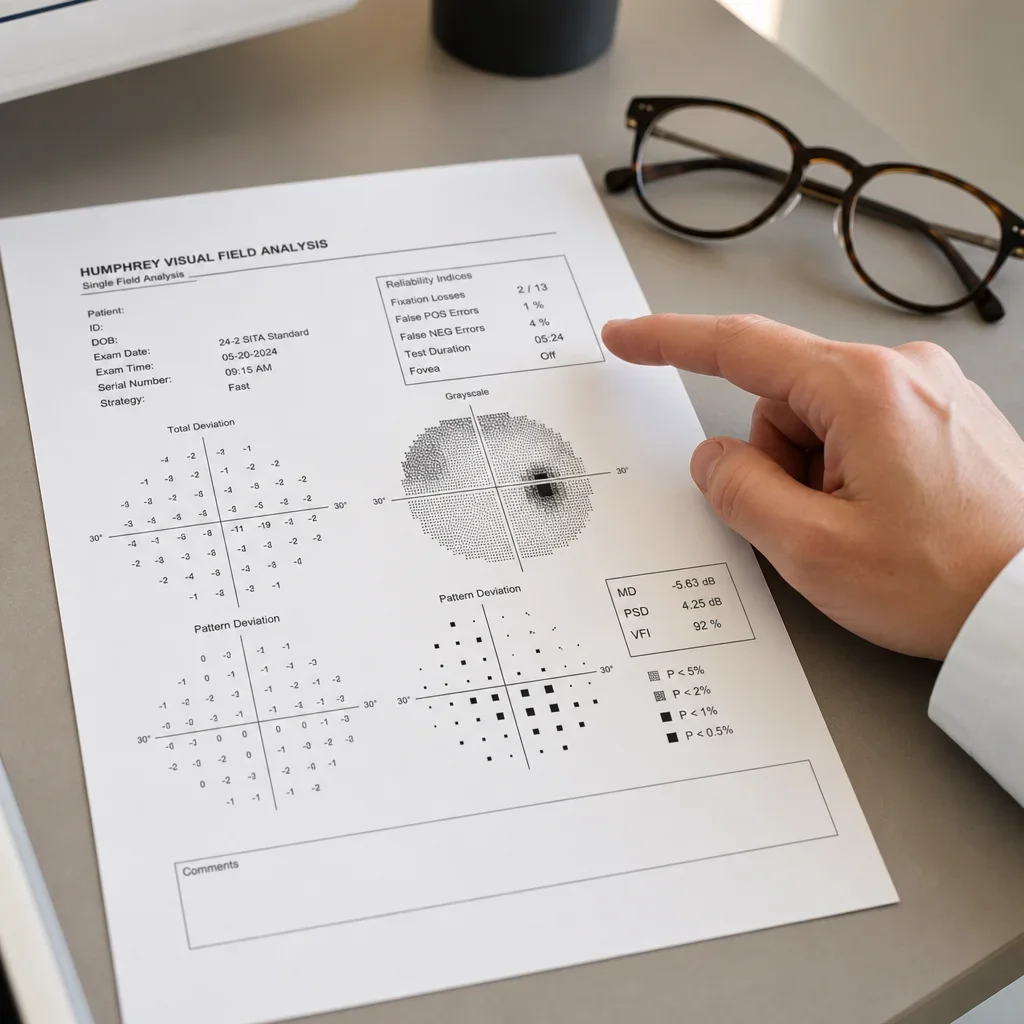
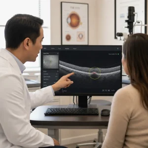
A Humphrey visual field report usually shows patient details, the eye tested, the program used, the test strategy, reliability information, and maps or values showing where responses were stronger or weaker. The printout is designed for clinical interpretation, but patients can still learn the basics.
The first things to look at on Humphrey visual field test results are whether the correct eye and test pattern were used and whether reliability was acceptable. Fixation losses suggest the patient may not have kept steady gaze, false positives suggest over-clicking, and false negatives may reflect fatigue, inattention, or real disease in the right context.
Grayscale plots can look dramatic, but they are not the most trustworthy part of Humphrey visual field interpretation. Clinicians care more about repeatability, pattern location, threshold data when available, reliability indices, and whether the defect makes anatomical sense.
Mean deviation can be a useful summary index on some visual field reports, but its value and trendability depend on the exact program and output. For a 60-4, you should not assume every printout supports the same trend analysis as a standard glaucoma follow-up field. Ask the clinic which summary measures are available on that machine.
Anderson’s criteria visual field language is part of glaucoma-style interpretation in some settings, but criteria created for standard central field analysis may not translate neatly to every wide peripheral program. That is another reason a 60-4 should be interpreted by the clinician who knows why it was ordered.
Common artifacts and why far-peripheral tests can be tricky
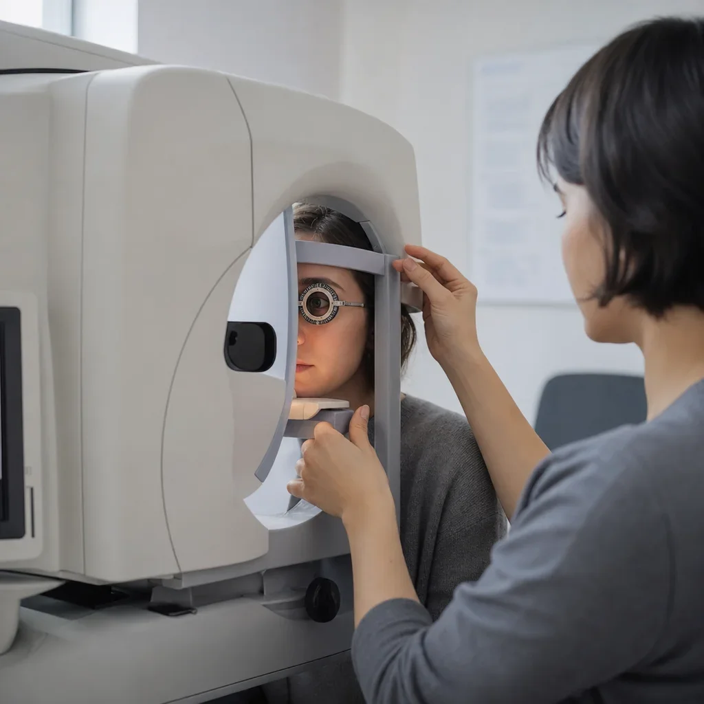
Far-peripheral field testing is more vulnerable to artifacts than central testing. Brow shadow, nose position, deep-set eyes, ptosis, dry eye, poor alignment, trial lens rim effects, fatigue, and misunderstanding the task can all create defects that are not true disease.
Facial contour matters because the more peripheral the light stimulus, the more likely it can be blocked by eyelids, brow, nose, or poor head position. This is one reason a suspicious peripheral defect may improve after better positioning or repeat testing.
The practical fixes are simple: keep the head steady, blink normally, ask for the instructions again if needed, mention dryness or discomfort, and let the staff reposition you if something feels off. If a defect only appears once and does not fit the exam, it may be artifact rather than pathology.
Peripheral testing is considered less common or more controversial in some settings because it can produce more noise and because standard central programs answer more routine clinical questions. Wider testing still has value, but only when it fits the reason for testing.
Pros and cons of the 60-4 visual field test
The biggest advantage of the 60-4 visual field test is wider peripheral assessment. That can help when symptoms, disease pattern, or documentation needs extend beyond the central field tested by 24-2 or 30-2.
| Pros | Cons |
|---|---|
| Assesses wider peripheral vision | More prone to artifact than central tests |
| May help explain tunnel vision or mobility complaints | Can feel longer or more tiring |
| Useful in selected retinal or neuro-ophthalmic cases | Less routine for standard glaucoma follow-up |
| May support documentation of field extent | Interpretation depends heavily on positioning and reliability |
The main trade-off is that wider does not always mean better. A test is only useful if it answers the right clinical question with enough reliability to act on.
How often the test may need to be repeated
A 60-4 may be repeated to confirm an abnormal finding, monitor a condition known to affect peripheral vision, document change over time, or check whether an odd result was caused by artifact. The interval depends on the disease being followed, symptoms, risk of progression, and how reliable the first result was.
A borderline or unexpected field is often repeated before major decisions are made. A one-time documentation need is different from long-term monitoring, and screening is different again from disease follow-up.
Cost, insurance, and medical necessity
Coverage for a Humphrey visual field test cost depends on medical necessity, the diagnosis being investigated, the clinic’s billing model, and your insurance plan. In Ontario, patients should confirm coverage with the clinic and their insurer because public and private coverage rules are not the same thing.
U.S. search results may mention CPT codes such as 92083, but that is U.S.-specific billing language and not Ontario billing advice. Patients in Waterloo or Kitchener should ask what the test is being billed as, whether direct insurance billing is available, and whether the plan requires a medical reason or referral.
We would not quote an out-of-pocket price without the clinic’s current fee schedule. Cost can differ based on whether testing is part of a broader exam, a diagnostic add-on, or a specialist referral workflow.
Driving, legal blindness, and other common misconceptions
A 60-4 result does not by itself decide whether someone is legally blind or allowed to drive. Those decisions depend on jurisdiction-specific rules and can involve both visual acuity and visual field findings.
Acuity numbers like 6/60 or 20/40 are not visual field numbers. If you are asking, “Can I drive with a 6-60 vision?” or “Is 60 eyesight legally blind?” the first step is to separate acuity from field testing, then check the actual Ontario standard rather than relying on forum answers.
A visual field test also does not answer treatment questions such as the newest glaucoma treatment in 2026. Treatment decisions depend on the diagnosis, severity, pressure control, imaging, medications, laser options, surgery discussions, and specialist guidance.
Who may need alternatives or special accommodations
Some patients need extra coaching or a different testing approach. That includes low-vision patients, children, people with limited mobility, patients with anxiety, and anyone who has trouble maintaining attention or understanding the instructions.
Not every patient produces a reliable standard automated field on the first try. Breaks, repeat explanation, modified positioning, and alternate methods may be more appropriate in selected cases.
When to book an eye exam or diagnostic visit
New side-vision symptoms, tunnel vision, unexplained visual changes, or glaucoma monitoring needs are good reasons to book an eye exam promptly. A visual field test is only one piece of the workup, so the right next step is usually a comprehensive assessment rather than trying to decode a printout alone.
If you are looking for an optometrist in Waterloo or need an eye exam in Waterloo after being advised to get visual field testing, ask the clinic what diagnostic testing is available, whether direct billing applies, and whether you need a general exam or a focused diagnostic visit. Premier Optical offers comprehensive eye exams, eyewear, and direct billing under one roof, but patients should confirm whether a specific visual field program is offered before booking.
FAQ
What is a 60/4 visual field?
It is a wide-field visual field testing pattern used to assess more peripheral vision than standard central tests. It measures where you detect light around you while you keep looking straight ahead.
What does the 60 4 visual field test measure?
It measures peripheral visual sensitivity, not visual acuity. In plain language, it checks how much of the surrounding field you can detect.
Is the 60-4 visual field test used for glaucoma?
Sometimes, yes, but it is usually not the default routine glaucoma test. Central patterns such as 24-2 are more commonly used for regular glaucoma monitoring.
What is the difference between 60-4 and 24-2 visual field testing?
A 24-2 is a central field test commonly used in glaucoma care. A 60-4 looks farther into the peripheral field and is used when the wider field matters more than dense central sampling.
What is a normal visual field test result?
A normal result is one that falls within expected limits for the patient’s age and is reliable enough to interpret. There is no single universal normal number for every visual field program.
What is an abnormal visual field?
An abnormal field may show repeatable missed areas, reduced sensitivity, asymmetry, or generalized depression. It can also be abnormal because the test was too unreliable to interpret confidently.
How long does a visual field test take?
Most automated visual field tests take about 5 to 15 minutes per eye depending on the program, pauses, and whether any section must be repeated.
How should I prepare for a Humphrey visual field test?
Bring your distance correction if the clinic told you to, arrive rested if possible, and expect to focus on a central target while pressing a button for lights you see. Keep looking straight ahead and do not chase the lights.
Can an eye doctor detect multiple sclerosis on a visual field test?
A visual field test can show findings that suggest optic nerve or neurologic issues, but it does not diagnose multiple sclerosis by itself. Further assessment is needed when a field pattern raises concern.
Will insurance cover a 60-4 visual field test?
Coverage depends on medical necessity, your diagnosis, and your insurance plan. Confirm with the clinic and your insurer because coverage rules differ.
Does a 60-4 visual field test determine if someone can drive?
No. Driving decisions depend on jurisdiction-specific standards and may consider more than one vision measure.
Is a visual field test the same as a vision chart test?
No. A chart test measures acuity. A visual field test measures the area around you where you can detect targets.
If your side vision feels off, the useful next step is not guessing from a printout. It is getting the right exam and the right test for the question.