Most people hear “slit lamp exam” and picture a separate hospital test. It usually is not. A slit lamp exam is a close-up part of a comprehensive eye exam where the optometrist uses a microscope and a narrow beam of light to examine the front of the eye, and sometimes deeper structures too. It is also called a slit lamp examination, a slit lamp eye exam, or biomicroscopy. It shows far more detail than a penlight check and helps us understand whether irritation, dryness, redness, blurred vision, injury, or routine changes need treatment, monitoring, or simply reassurance.
What Is a Slit Lamp Exam?
A slit lamp exam is a microscope-based eye check that lets the optometrist examine the eye layer by layer. The instrument combines binocular magnification with a bright, adjustable beam of light, which is why you may also hear terms like biomicroscopic examination, slit lamp biomicroscopy, or biomicroscopy eye examination.
A slit lamp eye examination is usually part of a full eye exam, not a separate visit. We see it used for routine screening, red eyes, dry eye symptoms, contact lens issues, injuries, and follow-up of known eye conditions. The exact findings are recorded by structure, not as one pass-or-fail score.
A slit exam, slit eye exam, or eye slit test all mean roughly the same thing in patient language. The formal idea is simple: the optometrist slit lamp helps the doctor look at tiny eye structures in much higher detail than a basic torch light exam.
What Does a Slit Lamp Exam Check?
A slit lamp assessment checks the eyelids, lashes, tear film, conjunctiva, sclera, cornea, anterior chamber, iris, pupil, and natural lens. Those are the front-of-eye structures most often viewed during a standard slit lamp evaluation.
A slit lamp examination can also help assess deeper structures when extra lenses or dilation are used. In those cases, the optometrist may examine the vitreous, optic nerve, macula, and retina as part of a broader eye health assessment rather than a front-surface-only check.
The test helps detect surface problems, inflammation, infection, injury, and age-related lens changes. It can also show whether symptoms like watering, burning, foreign body sensation, or light sensitivity are coming from the tear film, cornea, eyelids, or inside the eye.
Structure-by-structure: what the doctor is looking for
| Eye structure | What the doctor checks | What may stand out |
|---|---|---|
| Eyelids and lashes | Lid position, swelling, debris, lash direction | Blepharitis, styes, misdirected lashes |
| Tear film | Tear quality and stability | Dry eye, fast tear breakup |
| Conjunctiva | Redness, swelling, discharge, spots | Allergy, irritation, infection |
| Sclera | Colour and surface appearance | Inflammation or irritation |
| Cornea | Clarity, smooth surface, staining, injury | Abrasion, ulcer, dryness, foreign body |
| Anterior chamber | Inflammation, blood, pus, depth | Cells, flare, hyphema, hypopyon |
| Iris and pupil | Shape, symmetry, reaction | Inflammation, trauma-related changes |
| Lens | Clarity and colour changes | Cataract changes |
| Vitreous and retina* | Floaters, hemorrhage, retinal health | Assessed with added lenses and sometimes dilation |
*Viewed when needed with additional techniques, not every routine front-eye scan.
How a Slit Lamp Works
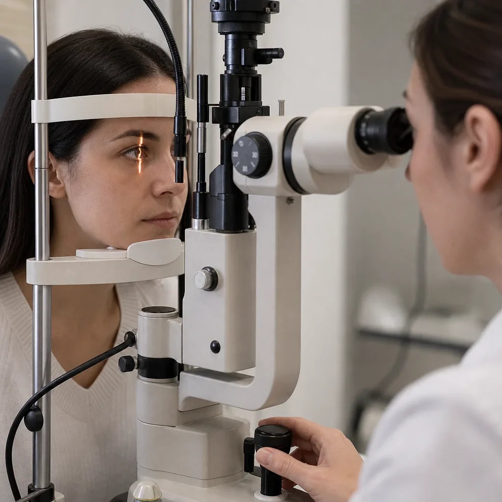
A slit lamp works by pairing a microscope with a thin beam of light. The microscope magnifies what the optometrist sees, and the “slit” of light creates a narrow optical section that helps show depth in transparent tissues like the cornea and lens.
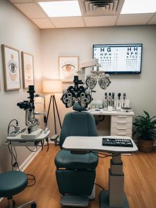
The slit lamp machine has a chin rest and forehead bar to keep your head steady. We ask patients to sit still, rest their chin, and look where directed, because tiny movements can blur what the doctor is trying to inspect.
The light can be changed in width, height, angle, brightness, and colour filter. That is why slit lamp illumination techniques matter. One setting may highlight the tear film, another may make corneal scratches easier to see, and another may help the doctor judge whether the lens is clear or cloudy.
The main slit lamp parts and functions are straightforward. The chin and forehead supports position you, the binocular microscope magnifies the view, and the adjustable beam reveals shape, depth, and clarity in different tissues.
What Happens During a Slit Lamp Eye Exam
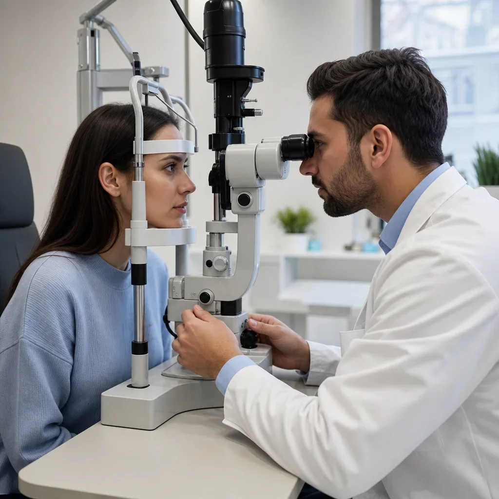
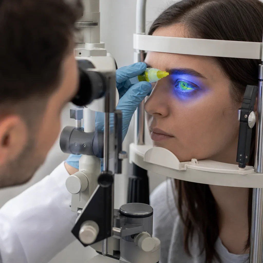
The slit lamp examination procedure is usually brief and direct. You sit at the instrument, place your chin on the rest and your forehead against the bar, and the optometrist lines up the light and microscope.
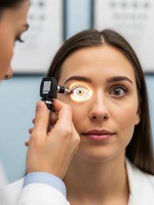
The doctor then examines the outer eye and front eye while asking you to look straight ahead or in a few directions. The bright beam may feel intense for a few seconds at a time, but the light exposure is brief and the test itself is not usually painful.
Fluorescein dye may be added if the doctor needs a better view of the tear film or corneal surface. This is common when someone has dryness, irritation, contact lens trouble, or a possible scratch or foreign body.
Dilation may be added when the optometrist needs a better look inside the eye. That is not automatic for every slit lamp eye exam, and it is one reason a slit lamp exam is not the same thing as a dilated eye exam.
A simple five-step version looks like this:
1. Position at the instrument. 2. Check lids, lashes, tears, and eye surface. 3. Scan the cornea, anterior chamber, iris, and lens. 4. Add fluorescein or other drops if needed. 5. Review findings and decide if more testing, treatment, or follow-up is needed.
Is a Slit Lamp Exam Painful or Dangerous?
A slit lamp exam is generally not painful. The usual complaint is bright light, not pain, and that light sensitivity tends to be brief unless the eye is already dry, inflamed, or injured.
The slit lamp itself does not damage healthy eyes when used properly in a routine clinical setting. The instrument shines light and magnifies structures. It does not touch the eye during a standard exam unless a separate contact-lens-based test is being done for another reason.
Drops can be the part you notice more. Fluorescein, anesthetic drops, or dilating drops may sting for a moment, and dilation can leave some people with temporary blur or light sensitivity afterward. The optometrist decides whether drops are needed based on your symptoms and eye health.
Some symptoms should not wait. Sudden vision loss, worsening eye pain, marked redness with light sensitivity, halos with nausea, or persistent symptoms after an eye injury need urgent eye assessment rather than a routine wait-and-see approach.
Do You Need to Prepare for a Slit Lamp Exam?
Most routine slit lamp examinations need little to no special preparation. Bringing the right things matters more than fasting, avoiding screens, or doing anything unusual beforehand.
Bring your current glasses, contact lenses, contact lens case, medication list, and insurance information if relevant. If you have a copy of an outside prescription or a recent eye history, bring that too.
Contact lenses may need to come out for part of the visit. That is especially common when the optometrist is checking the cornea, dry eye, surface irritation, or contact lens fit.
Heavy eye makeup can make the eyelid margin and tear film harder to assess. We usually tell patients to keep eye makeup light if they are coming in for redness, dryness, irritation, or a possible infection.
Medical details can change the exam plan. Tell the clinic about eye drops, medication allergies, pregnancy, recent eye surgery, diabetes, autoimmune conditions, recent injury, and any sudden change in vision.
Before-appointment checklist
- Bring your glasses and sunglasses.
- Bring contact lenses and your case.
- Bring a medication list or photos of labels.
- Mention allergies to drops or dyes.
- Mention pregnancy or breastfeeding.
- Mention recent injury or eye surgery.
- Mention new flashes, floaters, pain, or light sensitivity.
- Keep eye makeup minimal if the eye surface is being assessed.
Will Your Eyes Be Dilated, and Can You Drive Afterward?
A slit lamp exam and a dilated eye exam are not the same test. The slit lamp is the viewing instrument and exam method. Dilation is an added step that widens the pupil so the doctor can see more of the eye’s internal structures.
Not every patient needs dilation at every visit. The optometrist may recommend it for a better retinal view, diabetic screening, unexplained blurred vision, flashes or floaters, optic nerve assessment, or certain medical follow-ups.
Driving after a routine slit lamp exam is usually fine if no dilating drops were used and your vision feels normal. If you were dilated, driving may be uncomfortable or unsafe for some people because of glare, light sensitivity, and temporary blur.
Sunglasses help after dilation. We see this every bright winter day in Waterloo, when snow glare already makes light sensitivity worse. If you are sensitive to drops or have never been dilated before, arrange flexible transportation.
Does the Exam Use Fluorescein Dye?
A slit lamp exam sometimes uses fluorescein dye, but not always. Fluorescein is a yellow-orange dye that helps the doctor see the tear film and corneal surface more clearly under a blue light filter.
Fluorescein helps reveal corneal abrasions, dry spots, areas of irritation, contact lens-related surface changes, and some foreign bodies. In plain terms, it makes small surface defects stand out that can be hard to see in ordinary white light.
The dye may be instilled as a drop or applied with a small moistened strip. Either way, the effect is temporary, and the yellow colour does not stain the eye permanently.
Contact lenses usually need to be removed before fluorescein is used. If you wear lenses, bring your case so we can store them safely during the exam.
What Eye Conditions Can a Slit Lamp Exam Help Detect?
A slit lamp exam can help detect or document dry eye, blepharitis, conjunctivitis, corneal abrasions, corneal ulcers, foreign bodies, keratitis, anterior uveitis, cataract changes, and other visible front-of-eye problems. Those are among the most common slit lamp examination uses in everyday optometry.
At the lids and lashes, the doctor may see blepharitis, blocked oil glands, styes, or lashes rubbing the eye. Those findings can explain watering, burning, crusting, and irritation that people often mistake for “just allergies.”
At the cornea and tear film, the doctor may see dry eye patterns, abrasions, ulcers, contact lens irritation, or a tiny retained foreign body. This is one reason a slit eye test is so useful after something blows into the eye or after a bad contact lens day.
Inside the front of the eye, the doctor may look for inflammatory signs such as cells and flare, blood in the anterior chamber, or other findings that need urgent treatment or closer follow-up. The slit lamp is especially valuable here because depth matters, not just colour or redness.
At the lens, the doctor can often see cataract changes. A slit lamp can detect cataracts, but whether they are mild, moderate, or visually significant depends on the full exam and your symptoms, not one glance alone.
With added lenses and, in some cases, dilation, the same exam setup can help assess the vitreous, optic nerve, and retina. That broader exam may help identify diabetic eye changes, retinal problems, or optic nerve changes, but the slit lamp is still one tool among others, not a stand-alone answer for every condition.
An eye exam can also show signs that suggest broader health issues, but it does not by itself diagnose conditions like multiple sclerosis, high cholesterol, aneurysm, or autoimmune disease. If something unusual appears, the next step may be more testing, monitoring, or referral.
What Is a Normal Slit Lamp Exam?
A normal slit lamp exam means the structures viewed looked healthy for that visit and did not show obvious inflammation, injury, infection, or other visible abnormalities. Patients may also hear “normal slit lamp,” “normal slit lamp examination,” or “normal slit lamp exam” in chart notes or summaries.
A normal slit lamp exam often includes a clear cornea, a stable tear film, quiet conjunctiva, a deep and quiet anterior chamber, a round iris and pupil appearance, and a clear lens or age-appropriate lens changes. “Quiet” in this setting means no visible inflammatory activity in the chamber the doctor is viewing.
Normal does not mean every possible eye condition has been ruled out. Some problems need pressure testing, retinal examination, imaging, visual field testing, follow-up over time, or a different type of eye exam to confirm.
Results are usually documented by structure. That means your chart may say the cornea was clear, the anterior chamber was quiet, or the lens had mild cataract change, rather than giving one overall grade for the whole slit lamp evaluation.
Normal vs abnormal in plain English
| Finding style | What it usually means |
|---|---|
| Normal / clear / quiet | No obvious visible problem in that structure at that visit |
| Mild dry eye changes | Tear film or surface dryness was seen |
| Staining / fluorescein uptake | The eye surface has an area of disruption |
| Cataract changes | The natural lens is becoming cloudy |
| Cells and flare | Inflammatory signs were seen inside the eye |
| Foreign body | A small particle is present on or in the front eye tissues |
Common Findings and What They May Mean
Common slit lamp findings are descriptive terms, not final self-diagnoses. They tell the doctor what was seen and help decide whether you need treatment, monitoring, or referral.
Cells: tiny inflammatory cells floating in the anterior chamber. In plain English, this suggests irritation or inflammation inside the front of the eye.
Flare: a hazy protein appearance in the anterior chamber. In plain English, it can point to inflammation and is interpreted along with the rest of the exam.
Hyphema: blood in the anterior chamber. This can happen after trauma and needs professional assessment.
Hypopyon: a visible layer of inflammatory material or pus-like white cells in the anterior chamber. This is not a routine finding and needs urgent evaluation.
Keratic precipitates: inflammatory deposits on the back surface of the cornea. They can be seen in certain inflammatory eye conditions.
Fluorescein uptake: the dye highlighted an area on the cornea or conjunctiva. In plain English, the surface is disrupted there, as with a scratch, dry spot, or ulcer-related defect.
Abrasion: a scratch on the corneal surface. These are often painful and light-sensitive, but severity varies.
Ulcer: an infected or inflamed open defect on the cornea. This is more serious than a simple abrasion and needs prompt care.
Foreign body: a particle on the eye surface or embedded in the front tissues. Metal work, grinding, yard work, and windy days are common causes.
Cataract: clouding of the natural lens. A slit lamp can show cataract changes, but treatment decisions depend on how much vision is affected.
Abnormal findings can lead to several next steps. The optometrist may prescribe drops, recheck the eye, order further testing, dilate the pupils, refer you, or send you for urgent care depending on what was seen.
Routine vs Urgent: When a Slit Lamp Exam Is Needed Quickly
A slit lamp exam is routine in many comprehensive eye exams, but it also becomes urgent when symptoms suggest injury, infection, or internal inflammation. The instrument is useful in both settings. The urgency depends on symptoms and findings, not the machine itself.
Prompt assessment matters for sudden eye pain, marked redness with light sensitivity, a foreign body sensation after grinding or metal work, eye injury, sudden blurred vision, new flashes or floaters with a curtain-like shadow, or sudden vision loss. Those symptoms should not be left to a routine checkup weeks later.
More routine symptoms still deserve attention, but usually on a standard booking timeline. That includes gradual blur, mild dryness, itchy eyes, contact lens discomfort, and eyelid crusting that has not suddenly escalated.
In Waterloo and Kitchener, we see both patterns. Students wait too long with a painful contact lens problem, and parents wait through a weekend red eye hoping it settles. Some do. Some should have been seen sooner.
Urgent symptom callout
Seek prompt eye assessment for:
- sudden eye pain
- sudden vision loss or major blur
- redness with strong light sensitivity
- eye injury or chemical splash
- metal or debris in the eye
- flashes or floaters with a shadow or curtain
- severe symptoms after contact lens wear
Slit Lamp Exam vs Dilated Eye Exam, Woods Lamp, Tonometry, Gonioscopy, and Fundus Exam
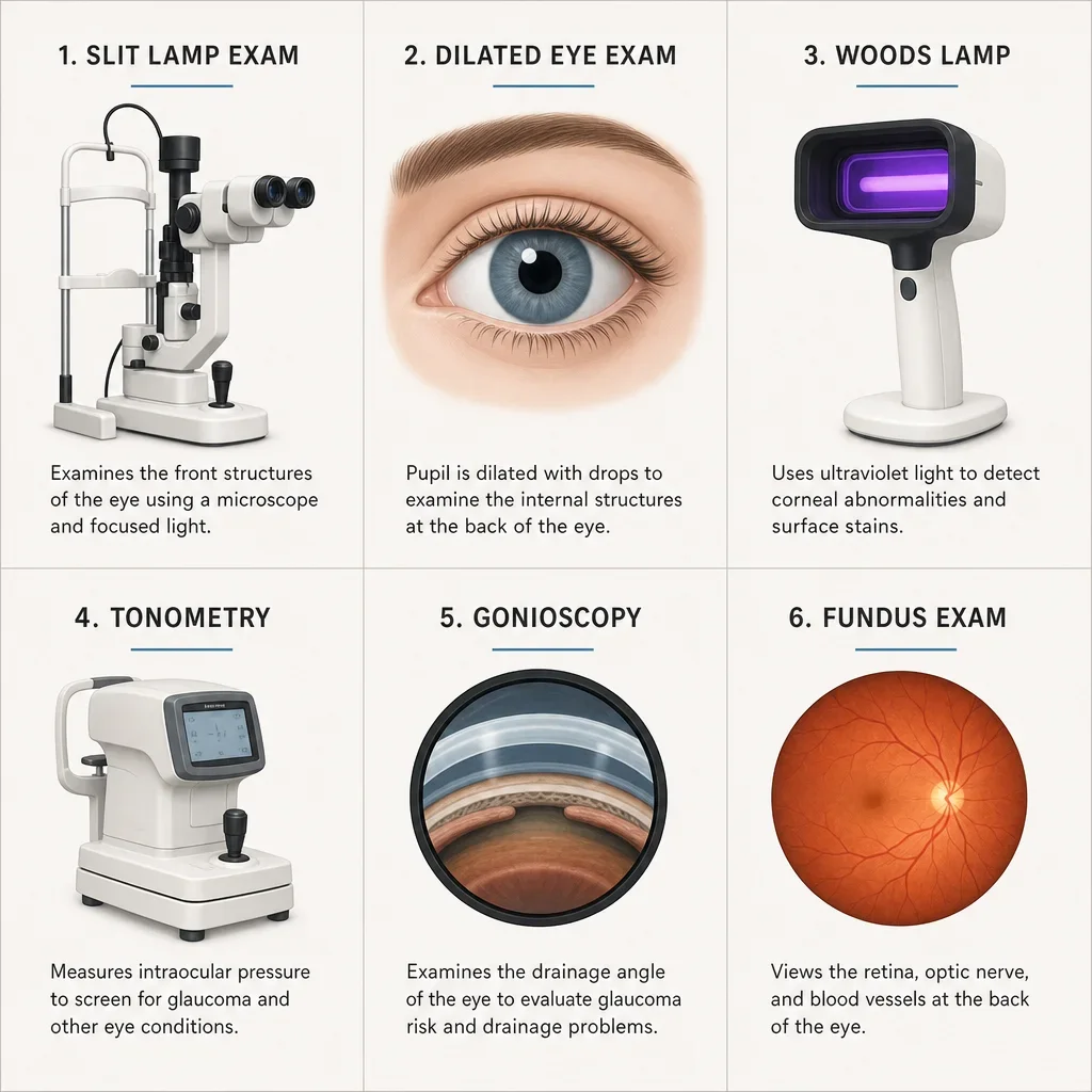
These tests are not interchangeable. Each one answers a different question, even when some are done in the same appointment.
| Test | What it checks | When it is used | Are drops typically needed? |
|---|---|---|---|
| Slit lamp exam | Front eye structures and, with added lenses, some deeper structures | Routine eye exams, red eye, dry eye, injury, follow-up | Not always |
| Dilated eye exam | Wider view of the retina, optic nerve, and internal eye | Retinal assessment, diabetes, flashes/floaters, fuller internal exam | Yes, for dilation |
| Woods lamp | Surface fluorescence with lower-detail screening | More limited surface screening, not a full substitute for slit lamp assessment | Sometimes dye is used |
| Tonometry | Intraocular pressure | Glaucoma screening and monitoring | Sometimes numbing drops, depending on method |
| Gonioscopy | Drainage angle of the eye | Glaucoma assessment, narrow-angle concern | Usually yes |
| Fundus exam | Retina, blood vessels, macula, optic nerve | Internal eye health assessment | Often dilation or special viewing methods |
A dilated eye exam is not the same as a slit lamp exam. Dilation is an added step. The slit lamp is the viewing system that can be used before and after dilation for different structures.
A Woods lamp is not the same as a slit lamp. In eye care, it is more limited and does not replace the detail of a slit lamp biomicroscopic examination.
Tonometry measures intraocular pressure. It does not inspect the cornea, lens, tear film, or anterior chamber the way a slit lamp does. Both tests may happen in one comprehensive exam because they answer different questions.
Gonioscopy is a specialized angle exam done with a lens placed on the eye after anesthetic drops. It uses the slit lamp as a platform, but it is not the same as a standard slit lamp scan.
A fundus exam focuses on the back of the eye, especially the retina and optic nerve. That may be done through dilation or with additional lenses during slit lamp fundoscopy, depending on what the doctor needs to assess.
Questions Patients Often Ask Before the Appointment
Contact lenses may need to be removed, especially if the cornea, tear film, or contact lens fit is part of the reason for the visit. Bring your case even if you expect to wear the lenses in.
Light eye makeup is usually fine for a routine visit, but heavy liner, false lashes, or thick mascara can make it harder to assess the lid margin and tear film accurately.
Mention allergies, pregnancy, breastfeeding, and all current eye drops before the exam. Those details can affect which drops the optometrist chooses and whether dilation or other testing is appropriate.
A slit lamp can help detect cataract changes and dry eye signs. It can also show inflammation, abrasions, ulcers, and foreign bodies. It does not, by itself, confirm every eye disease or every cause of blurred vision.
A slit lamp can contribute to glaucoma assessment, but it does not replace pressure testing, optic nerve evaluation, visual field testing, or other glaucoma workup. In practice, glaucoma care uses several findings together.
After the Exam: What to Expect Next
Most people can return to normal activity right after a routine slit lamp exam if no dilating drops were used and vision feels normal. There is usually no recovery period from the light itself.
If fluorescein was used, any yellow tint tears leave behind is temporary. If your contact lenses were removed, ask when it is safe to put them back in, because the answer depends on what the doctor found on the cornea and what drops were used.
If dilation was used, expect more light sensitivity and possible blur for a while afterward. Bring sunglasses, avoid rushing back into bright light, and be cautious with driving, reading, or screen-heavy tasks until your vision feels steady.
If an abnormal finding was seen, follow-up may range from routine monitoring to urgent treatment. We explain the time, cost, and next step as clearly as we can, but the exact plan depends on the structure involved and the severity the optometrist sees.
After-exam checklist
- Wear sunglasses if you were dilated.
- Do not force contact lenses back in without asking.
- Use prescribed drops exactly as directed.
- Watch for worsening pain, redness, or vision.
- Book the recommended follow-up, especially for injury or inflammation.
What to Expect at Premier Optical in Waterloo
At Premier Optical, a slit lamp exam may be part of a comprehensive eye exam with a licensed optometrist in Waterloo. If the doctor finds something that affects your prescription, lens comfort, or eyewear use, we can usually handle the next step under the same roof, from exam notes to frame fitting to lenses cut in our in-house lab.
That all-in-one setup matters when symptoms lead to practical questions. If the exam shows dry eye, contact lens irritation, or a prescription change, we can explain what that means for lenses, coatings, screen use, and follow-up without sending you across town. We also direct-bill many insurance plans where eligible.
If you have dryness, redness, blurred vision, a contact lens problem, or you are simply due for a checkup, booking an eye exam is the right next step. We would need to see your eyes, symptoms, and prescription history to say anything more specific.
FAQ
What is a slit lamp exam?
A slit lamp exam is a close-up eye examination done with a microscope and a narrow beam of light. It helps the optometrist examine the eyelids, tear film, cornea, anterior chamber, iris, and lens in detail.
Is the slit lamp exam painful?
Usually no. The light can feel bright, and irritated eyes may feel more sensitive, but the exam itself is generally not painful.
What does a slit lamp exam detect?
It can help detect dry eye, blepharitis, conjunctivitis, corneal scratches, foreign bodies, inflammation, and cataract changes. With added lenses or dilation, it can also contribute to deeper eye assessment.
What is a normal slit lamp exam?
A normal slit lamp exam means the visible structures looked healthy and free of obvious injury, inflammation, or other visible abnormalities at that visit. It does not rule out every possible eye condition.
Will my eyes be dilated for a slit lamp exam?
Not always. Dilation is only used when the optometrist needs a better view of internal eye structures or needs more information from the exam.
Is a slit lamp exam the same as a dilated eye exam?
No. A slit lamp exam is the viewing method. Dilation is an added step that may be used during a broader internal eye exam.
Can I drive after a slit lamp exam?
Usually yes if you were not dilated and your vision feels normal. If dilating drops were used, driving may be uncomfortable or unsafe for some people.
Does a slit lamp exam use fluorescein dye?
Sometimes. Fluorescein dye is used when the doctor needs a clearer view of the corneal surface or tear film.
What should I avoid before a slit lamp exam?
There is usually little to avoid. Heavy eye makeup, forgotten medication details, and coming in with contact lenses but no case are the main practical problems we see.
Can a slit lamp diagnose cataracts or dry eye?
It can show cataract changes and dry eye signs clearly, but the full diagnosis and treatment plan depend on the complete exam and your symptoms.
Can a slit lamp detect glaucoma?
It can contribute to glaucoma assessment, but glaucoma is not diagnosed by slit lamp findings alone. Pressure testing, optic nerve evaluation, and other tests may also be needed.
Can a slit lamp damage your eyes?
Not in routine clinical use. The bright light may be uncomfortable, but the slit lamp itself does not damage the eyes when properly used.
If you are still unsure whether your symptoms sound routine or urgent, call before you wait it out. A short conversation usually tells us whether you need the next available eye exam in Waterloo or faster care.