What Is an OCT Eye Exam? OCT, Visual Field Tests, and Eye Doctors Explained
Your optometrist says you need an “OCT scan” and a “visual field test.” You nod like you understand. But as soon as you sit back down in the waiting room, the same questions hit: What are these tests? Are they going to hurt? And why can’t a regular eye exam catch whatever they’re looking for?
These are two of the most important diagnostic tests in modern eye care — and most people have never heard of either one until they’re told they need one. This guide explains what each test does, what it detects, what happens during the procedure, and why your eye doctor may recommend them as part of a comprehensive exam.
First: What Do You Call an Eye Doctor?
Before diving into the tests themselves, let’s clear up a question that catches more people than you’d expect: what’s the difference between the types of eye doctors?
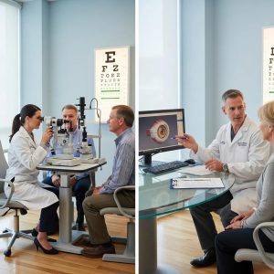
An optometrist (Doctor of Optometry, or OD) is the eye care professional most people see for their regular eye exams. Optometrists perform comprehensive eye exams, prescribe glasses and contact lenses, diagnose common eye diseases, and manage conditions like dry eye, glaucoma, and diabetic eye disease. In Ontario, optometrists are regulated by the College of Optometrists of Ontario. They’re the ones performing OCT scans and visual field tests in the clinic.
An ophthalmologist is a medical doctor (MD) who specializes in eye and vision care. Ophthalmologists do everything an optometrist does, plus they perform eye surgery — cataract removal, LASIK, retinal repairs, and other procedures. If your optometrist finds something during an OCT or visual field test that requires surgical intervention, they’ll refer you to an ophthalmologist.
An optician is the professional who fits and dispenses your glasses and contact lenses based on the prescription written by your optometrist or ophthalmologist. They don’t examine your eyes or diagnose conditions.
| Optometrist (OD) | Ophthalmologist (MD) | Optician | |
| Eye exams | Yes | Yes | No |
| Prescribes glasses/contacts | Yes | Yes | No (dispenses only) |
| Diagnoses eye diseases | Yes | Yes | No |
| OCT and visual field tests | Yes | Yes | No |
| Eye surgery | No | Yes | No |
| Fits and adjusts eyewear | Sometimes | No | Yes |
What Is an OCT Eye Exam?
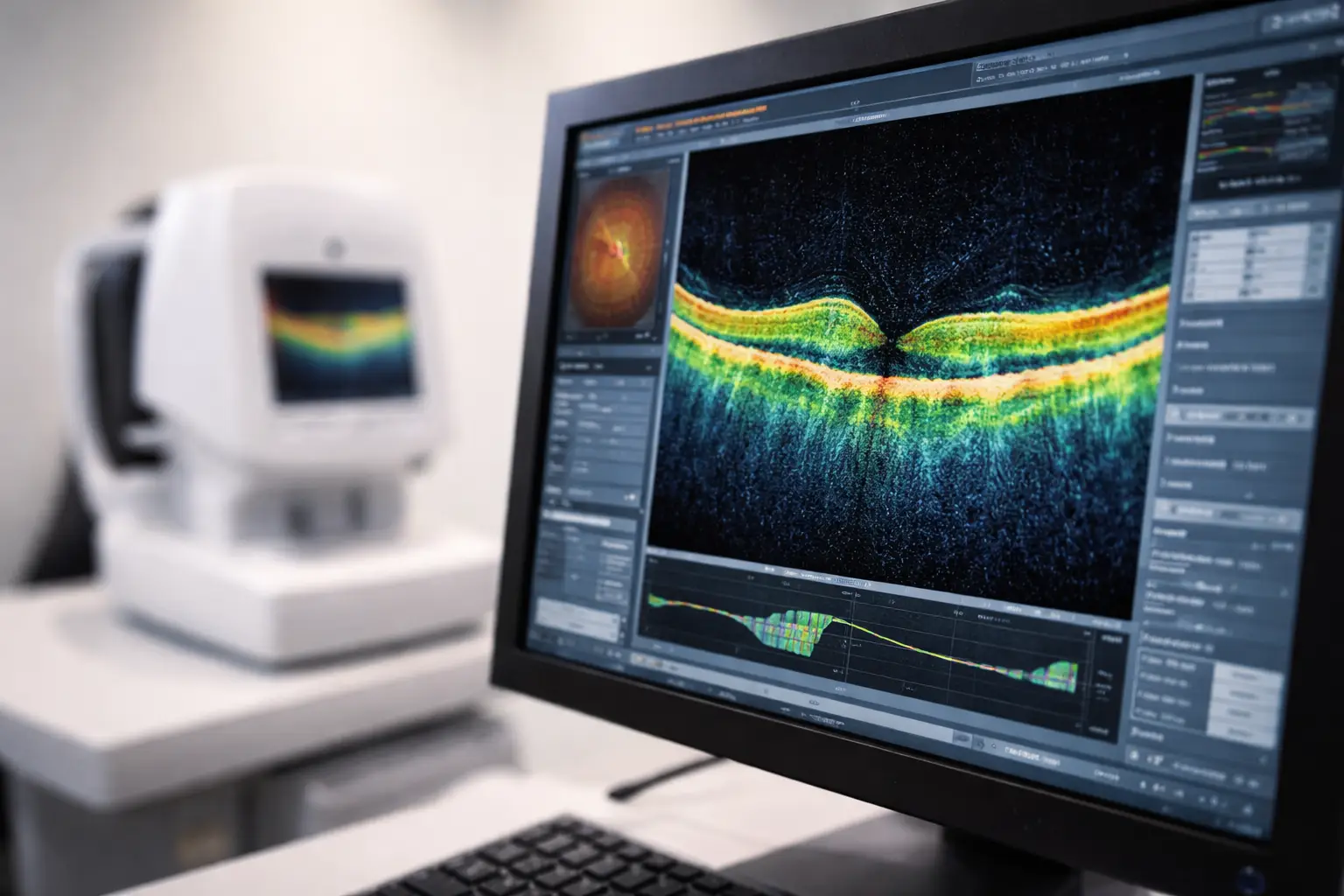
OCT stands for Optical Coherence Tomography. It’s a non-invasive imaging test that uses infrared light waves to create highly detailed, cross-sectional images of the inside of your eye — specifically the retina (the thin tissue at the back of the eye that processes light) and the optic nerve (which sends visual signals to your brain).
Think of it like an ultrasound, but using light instead of sound. The result is a 3D map of your retina that shows each individual layer in microscopic detail. Your optometrist can measure the thickness of these layers, compare them against normal ranges, and spot abnormalities that would be invisible during a standard eye exam — even one that includes looking at the back of your eye with a light.
What Does an OCT Scan Detect?
An OCT scan is one of the most powerful tools available for detecting and monitoring eye diseases that damage the retina and optic nerve. Conditions it helps diagnose include:
- Glaucoma — by measuring the thickness of the retinal nerve fibre layer (RNFL) around the optic nerve. Thinning of this layer is one of the earliest signs of glaucoma, often appearing years before you notice any vision loss. OCT can detect glaucoma up to 4 years earlier than traditional methods.
- Age-related macular degeneration (AMD) — by showing fluid buildup, drusen deposits, or thinning in the macula (the central part of the retina responsible for sharp, detailed vision). OCT can also distinguish between dry AMD and wet AMD, which require different treatments.
- Diabetic retinopathy and diabetic macular edema — by revealing swelling, leaking blood vessels, and abnormal tissue growth in the retina caused by diabetes.
- Macular holes and epiretinal membranes — structural defects in the retina that can cause distorted or blurred central vision.
- Retinal detachment — by showing the retina pulling away from the tissue beneath it, which is a medical emergency.
- Optic nerve disorders — including optic neuritis (inflammation of the optic nerve, sometimes associated with multiple sclerosis).
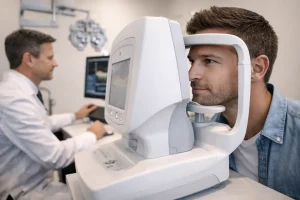
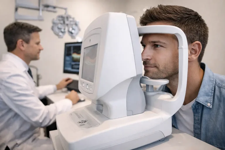
What Happens During an OCT Scan?
The entire test takes about 5 to 10 minutes. Here’s exactly what to expect:
- Your optometrist may use dilating eye drops to widen your pupils, though this isn’t always necessary for OCT.
- You sit in front of the scanner and rest your chin on a small support to keep your head still.
- You focus on a green target light inside the machine.
- The scanner takes images of each eye, one at a time. You may see a red line moving across your vision. Nothing touches your eye.
- You won’t feel any discomfort. The test is completely painless and non-invasive.
If dilating drops are used, your vision will be blurry and light-sensitive for a few hours afterward. Bring sunglasses to your appointment.
What Is a Visual Field Test?
A visual field test (also called a visual field exam or perimetry) measures your entire range of vision — not just what you see directly in front of you, but your peripheral (side) vision as well. While an OCT shows the physical structure of your retina, a visual field test measures how well your retina is actually functioning. The two tests are complementary: one looks at the anatomy, the other tests the performance.
What Does a Visual Field Test Detect?
A visual field exam is primarily used to detect and monitor:
- Glaucoma — the most common reason for visual field testing. Glaucoma typically causes blind spots in your peripheral vision first, then gradually narrows your visual field toward the centre. Most people don’t notice peripheral vision loss until it’s significant, which is why testing is so important.
- Neurological conditions — including brain tumours, strokes, or optic nerve damage. These conditions can cause very specific patterns of vision loss (for example, losing the same side of vision in both eyes) that point directly to the location of the problem in the brain.
- Retinal conditions — including retinitis pigmentosa and retinal detachment, which cause predictable patterns of peripheral vision loss.
- Monitoring treatment effectiveness — if you’re already being treated for glaucoma, your optometrist uses visual field tests to track whether the treatment is holding your vision stable or if your field is continuing to shrink.

What Happens During a Visual Field Test?
A standard automated visual field test takes about 5 to 10 minutes per eye. Here’s the procedure:
- You sit in front of a bowl-shaped instrument and rest your chin on the support.
- One eye is covered with a patch. You focus on a fixed point in the centre of the bowl with the other eye.
- Small lights flash at different locations and intensities across the bowl. Each time you see a flash, you press a button.
- The machine maps which areas of your visual field responded to the light and which didn’t. Missing a few flashes is completely normal — the test is designed to push the limits of your peripheral vision.
- The test is then repeated for the other eye.
The result is a printed map showing the sensitivity of every part of your visual field. Dark spots indicate areas where your vision is reduced or absent. Your optometrist compares this map against previous tests to track any changes over time.
OCT vs Visual Field Test: How They Work Together
These two tests answer different but equally important questions about your eye health:
| OCT Scan | Visual Field Test | |
| What it measures | Physical structure of the retina and optic nerve | Functional performance of your vision across your entire field |
| What it uses | Infrared light waves (non-invasive imaging) | Flashing lights you respond to with a button |
| Best at detecting | Structural thinning, fluid, swelling, deposits | Blind spots, peripheral vision loss, field narrowing |
| Key condition | Macular degeneration, diabetic eye disease | Glaucoma, neurological conditions |
| Time | 5–10 minutes total | 5–10 minutes per eye |
| Pain | None | None |
For conditions like glaucoma, your optometrist typically uses both tests together. The OCT shows whether the nerve fibre layer is physically thinning. The visual field test shows whether that thinning has started to affect your actual vision. Sometimes the OCT catches structural damage years before the visual field test shows any functional loss — which is exactly why early detection matters.

When Should You Get an OCT or Visual Field Test?
Not every patient needs these tests at every exam. Your optometrist will recommend them based on your specific risk factors. You’re more likely to need an OCT and/or visual field test if:
- You’re over 40 — the risk of glaucoma, macular degeneration, and other age-related conditions rises significantly after this age
- You have diabetes — diabetic retinopathy is one of the leading causes of vision loss in working-age adults, and OCT is the gold standard for monitoring it
- You have a family history of glaucoma or macular degeneration — genetics play a major role in both conditions
- You’re of African, East Asian, or Hispanic descent — these populations have higher rates of certain types of glaucoma
- You have high blood pressure or cardiovascular disease — both can damage retinal blood vessels
- You’re very nearsighted (high myopia) — severe nearsightedness stretches the retina and increases the risk of retinal tears, detachment, and glaucoma
- Your optometrist noticed something during your standard exam — like cupping of the optic nerve or changes in your retinal appearance — that needs closer investigation
In Ontario, adults aged 20 to 64 are not covered by OHIP for routine eye exams, but children and seniors (65+) are covered every 12 to 18 months depending on conditions. However, OHIP does cover medically necessary assessments — meaning if your optometrist identifies a condition that requires monitoring through OCT or visual field testing, those tests may be covered. Premier Optical in Waterloo offers direct billing for major insurance providers, university benefits plans, and corporate vision care — so your out-of-pocket cost for these advanced diagnostic tests is often minimal or zero.